Journal Information
Share
Share
Download PDF
More article options
Images in Clinical Hematology
Full text access
Available online 15 April 2024
Plasmodium ovale gametocytes in peripheral blood
Visits
85
Angela Lozano-Ochoaa,
Corresponding author
angela.lozanoochoa@osakidetza.eus

Corresponding author at: Hematology Department, Cruces University Hospital, Biscay, Spain.
, Verónica Roldán Galiachoa, Leyre Cristina Jiménez Rosb, Raquel Pérez Garayb, Juan Carlos García-Ruiza
a Hematology Department, Cruces University Hospital, Biscay, Spain
b Clinical Laboratory Department, Cruces University Hospital, Biscay, Spain
This item has received
Received 18 September 2023. Accepted 09 November 2023
Article information
Full Text
Bibliography
Download PDF
Statistics
Figures (1)
Full Text

A 54-year-old woman attended the emergency room with history of fever and myalgia. She had arrived from the Central African Republic one month previously. On admission, laboratory data showed: hemoglobin 70 g/L, platelets 41 × 109/L, leukocytes 0.76 × 10⁹/L, lactate dehydrogenase 330 U/L [normal: 0–245 U/L], total bilirubin 1.4 mg/dL and C-reactive protein 53.13 mg/L [normal: 0–11 mg/L].

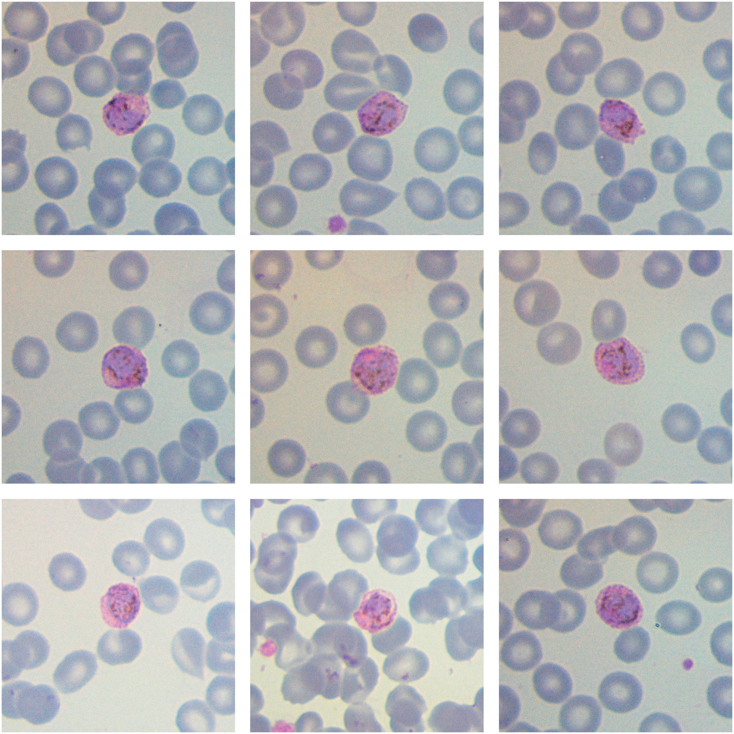
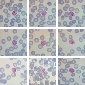
Rapid diagnostic tests for malaria as well as thick film performed upon arrival were negative. A peripheral blood film stained with May-Grünwald and Giemsa showed slightly enlarged red blood cells covered with Schüffner's dots and prominent brown pigment (hemozoin), suggesting infection by plasmodium at the gametocyte stage. The results obtained by quantitative polymerase chain reaction (qPCR) later on confirmed infection by Plasmodium ovale.

Malaria is a parasite infection caused by Plasmodium spp. Traditionally considered the rarest species, in recent years a rise in cases associated with P. ovale has been observed. Trophozoites are the most common morphological finding however, after a long incubation period of this species, gametocytes may be found. 1 Stippling is more pronounced and tends to darken the red blood cell. As P. ovale exclusively infects reticulocytes, the parasite density is typically low.2P. ovale is endemic in western Africa and the Southwest Pacific. The increase in migration flows underscores the need for an early microscopic identification of this infection, in order to achieve a swift diagnosis and guarantee correct treatment.

References
[1]
W.E. Collins, G.M Jeffery.
Plasmodium ovale: parasite and disease.
Clin Microbiol Rev, 18 (2005), pp. 570-581
[2]
M.L Wilson.
Laboratory diagnosis of malaria: conventional and rapid diagnostic methods.
Arch Pathol Lab Med, 137 (2013), pp. 805-811
Copyright © 2024. Associação Brasileira de Hematologia, Hemoterapia e Terapia Celular
Idiomas
Hematology, Transfusion and Cell Therapy
Article options
Tools