
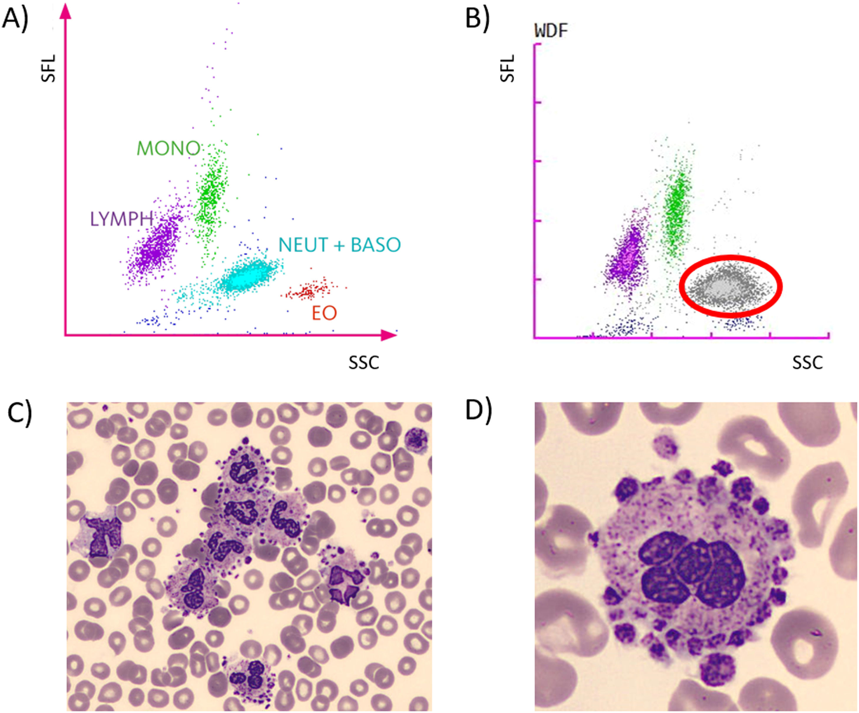
We here present a case of massive Platelet Sattlitism (PS) around neutrophils in a 91-year old man with a normal thrombocyte count, detected through an incidental abnormal white blood cell scattergram flag on the Sysmex XN—L550 cell counter (Figure 1a-b).1 Using the basic principles of flowcytometry, a scattergram depicts the blood cell formula based on the sideward scatter (SSC; X-axis) and the side fluorescence light (SFL; Y-axis). The SSC represents the size of the cell, the SFL represents its metabolic acitivty. Compared to a normal white blood cell scattergram (Figure 1a) the scattergram of the current case showed an increased SSC and FSL in the neutrophil cloud (Figure 1b). The SSC rise can be ascribed to sticking of the platelets to neutrophils, resulting in larger cells; the FSL rise can be ascribed tot he chronic inflammatory state of the patient (he was under treatment for prostate carcinoma). Since platelets stick to neutrophils in vitro, but not in vivo in patients with PS, it is often suggested as a possible cause of pseudothrombocytopenia.1
. Abnormal White Blood Cell scattergram flag on the Sysmex Cell counter; the grey cloud with red circle represents the abnormal white blood cells (b). Platelet sattelitism with clustering of neutrophils (c). Close-up of a neutrophil surrounded by platelets (d). SFL: Side Fluorescence Light; SSC: Sideward Scatter; MONO: monocytes; LYMPH: lymphocytes; NEUT + BASO: Neutrophils + Basophils; EO: Eosinophils.")
Incidental finding of platelet sattelitism upon blood smear review. Normal peripheral blood scattergram (A). Abnormal White Blood Cell scattergram flag on the Sysmex Cell counter; the grey cloud with red circle represents the abnormal white blood cells (b). Platelet sattelitism with clustering of neutrophils (c). Close-up of a neutrophil surrounded by platelets (d). SFL: Side Fluorescence Light; SSC: Sideward Scatter; MONO: monocytes; LYMPH: lymphocytes; NEUT + BASO: Neutrophils + Basophils; EO: Eosinophils.
PS was not observed around other cell types (Figure 1c and 1d). Retrospective review of all blood smears performed for this patient revealed the presence of PS in a sample dating from five monhts earlier as well.
Hematologists should be aware that PS can persist for several months and may also occur in patients with normal platelet counts.


