
Neoplastic diseases affect about 1:1000 women during pregnancy.1 This incidence varies according to region, age and the presence or absence of pre-invasive diseases.2
Lymphoma is the fourth most diagnosed cancer in this period, behind breast tumors, cervical cancer and melanoma.3 Its frequency has risen with the tendency in modern society of postponing pregnancy.4
The most common lymphoma in pregnant patients is Hodgkin's lymphoma, since its incidence spike coincides with the female fertile age. Any link between non-Hodgkin lymphoma and pregnancy has still not been elucidated, with few case reports being found in the medical literature worldwide.3,4
Burkitt's lymphoma is a very aggressive germinal B-cell lymphoma, frequently presenting extranodal sites. It is the second most common lymphoma in children, but it is very rare in adults. Three clinical variants are known: endemic, sporadic and immunodefficiency-related.5
In the sporadic form, the primary location of the disease involves the abdomen in 80% and the mandible in 14% of patients. In the endemic form, the mandibular and maxillary are involved in 60% of patients, abdominal commitment in 58% of cases, followed by the central and paraspinal nervous systems. An important geographic difference between these two forms is the association of Epstein–Barr virus (EBV) with the endemic form. However, only 15–20% of Burkitt's lymphomas seen in Europe and in the USA are associated with EBV. The frequency of this association in South America seems intermediate and, in Brazil, a frequency of 70% for EBV associated with Burkitt's lymphoma has been reported.5
Burkitt's lymphoma was the first identified neoplasm whose pathogenesis involved a chromosomal translocation as demonstrated by molecular biology. This mutation involves a translocation between the distal part of the long arm of chromosome 8 and chromosome 14 [t(8;14) (q24;q32)]. As a result of this rearrangement, the myc proto-oncogene in chromosome 8 is translocated to the immunoglobulin heavy chain locus in chromosome 14.6
Case reportA 25-year-old female patient born in Santa Rita, state of Paraíba consulted with a slight stinging abdominal pain in the left hypochondriac and lumbar regions, associated with diarrhea and melena for three months. The result of a b-HCG test, performed because of amenorrhea, was positive. The obstetric ultrasonography (USG) revealed a four-week two-day pregnancy. After one month, there was an increase in the pain, associated with nausea and weight loss. A new obstetric USG, performed during prenatal care, showed a morphologically normal fetus (12 weeks), in addition to right colon thickening in the mother, with an extensive (5.0cm×3.0cm) vascularized hypoechogenic formation next to the hepatic angle. The patient was asked to urgently seek a gastroenterologist.
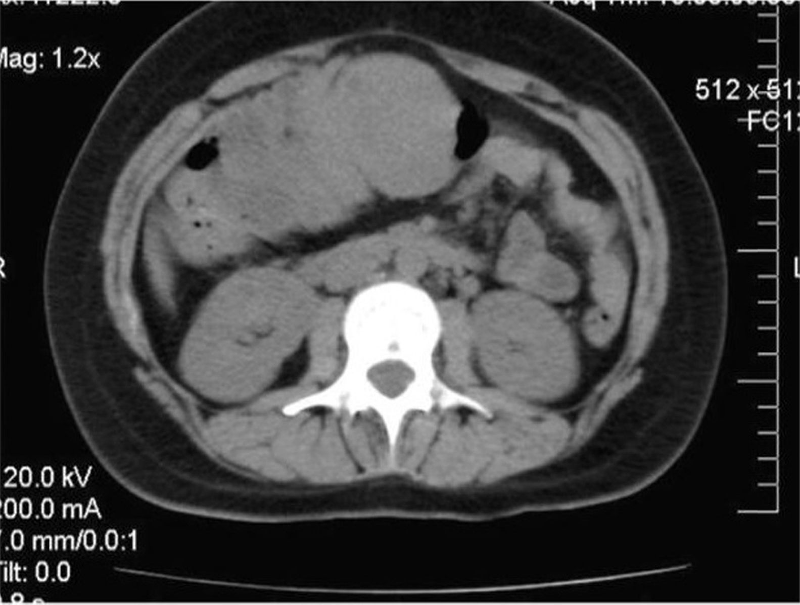
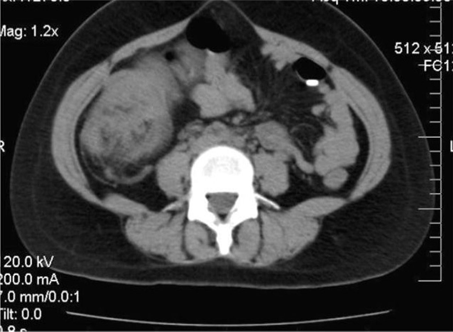
With significant worsening of the abdominal pain and acute exacerbation of the condition, the patient went to the Hospital Universitário Lauro Wanderley, where she was admitted to perform more tests. A computer tomography showed a soft tissue mass in the upper third part of the abdomen, with air-fluid level, and dilation of anterior intestinal loops with partial obstruction (Figures 1 and 2).


A further abdominal USG revealed an extensive (10.6cm×5.4cm) echo-complex mass in the right hypochondrial region, with a heterogeneous aspect (solid/cystic). A colonoscopy revealed a vegetative lesion of about 15cm occupying around 90% of the intestinal lumen.
The patient was submitted to enterectomy and typhlectomy, in addition to right segmental colectomy with primary latero-lateral ileocolonic anastomosis. Surgery was completed without complications and the patient evolved well after the procedure, without complaints of bleeding or pain in the lower abdomen. An evaluation of the fetal was made confirming preserved vitality. The patient was prescribed natural progesterone (400mg/day) in order to keep the uterus quiescent.
The mass removed during surgery was submitted to anatomopathological evaluation revealing medium and large cell non-Hodgkin lymphoma. Immunohistochemistry identified features of Burkitt's lymphoma. After discharge, the patient was referred for onco-hematological and obstetric care.
The staging of the lymphoma revealed isolated abdominal disease, with no bone marrow or cerebrospinal fluid involvement; it was classified as Murphy II-B. A 21-day regimen of rituximab IV (375mg/m2 – Day 1), cyclophosphamide IV (500mg/m2 per day – Day 1–3), vincristine IV (2mg – Day 1 and Day 8), doxorubicin IV (60mg/m2 – Day 1), oral prednisone (60mg/m2 – Day 1–5) (COPAD-R) was started following the LMB-96 protocol. Moreover, prophylaxis of the central nervous system was carried out using intrathecal chemotherapy: dexamethasone (2mg) and methotrexate (12mg – Day 2 and Day 5).7–9
The patient was advised about the aggressiveness of the condition and the possible risks to the fetus during chemotherapy, but chose not to interrupt pregnancy. The patient had a good evolution with clinical improvement.
During obstetric care, another morphological obstetric USG revealed a non-ectopic pregnancy with an echographic age of 19.1 weeks – in addition to an abdominal USG, which showed a solid abdominal mass (8.6cm×10.8cm) in a medial and slightly left position, which was heterogeneous with irregular edges. An USG performed the following week showed a non-ectopic pregnancy with echographic age of 20.3 weeks and preserved fetal morphology, and a solid abdominal mass (6.6cm×3.6cm×5.0cm), identical to the one previously described, indicating that there was a decrease in size after the beginning of chemotherapy.
After four cycles of chemotherapy, the patient evolved with a new acute abdominal condition. Laparotomy showed a perforation in the proximal jejunum, and so another enterectomy and urgent caesarian section (33rd week of pregnancy) were performed. A healthy female child of 1815g was delivered. The segment removed in the second enterectomy was sent for anatomopathological study, which showed no disease. A computer tomography after birth was normal.
The patient in this study died about three months after the date of birth, the victim of sudden death with no evidence of active oncological disease. The child remains healthy.
DiscussionBurkitt's lymphoma, although very aggressive, is highly treatable.10 The objective of treatment is to eliminate the largest number of malignant cells possible and induce complete remission, in other words, the disappearance of all evidence of the disease. Patients with aggressive and fast growing lymphomas are frequently treated with combined chemotherapy. Intensive chemotherapy with multiple drugs may be very effective against aggressive lymphomas. The prognosis of Burkitt's lymphoma depends on the extent of the disease and the time interval between the first complaints and diagnosis. The disease-free survival rate is between 75% and 85% in adults.6
There is no unique option for chemotherapeutic treatment of this kind of lymphoma. Today, the chemotherapeutic treatment consists in short-duration intensive regimens that include agents such as methotrexate, cyclophosphamide, vincristine, doxorubicin and prednisone. Methotrexate is used in most of the big centers, but it is highly toxic and is responsible for most of the risk during pregnancy. The treatment is complemented with prophylaxis for infections. Relapse sometimes occurs during the first year, and is a particularly poor prognostic sign.11
The treatment of a pregnant woman with cancer is always a delicate decision, since it involves risks for the mother and child, the wishes of the patient and the opinions of the oncologist, the obstetrician and the neonatologist. The potential effects of antineoplastic agents to the fetus include immediate effects, such as abortion and teratogenesis with specific damage to one or more organs, and late effects, such as delay in growth and gonadal dysfunction.1
The choice of the COPAD-R regimen following the LMB-96 protocol,7–9 aiming at greater practicality with fewer risks for the patient, was very successful, ensuring remission of the disease and completion of the pregnancy.
The rare occurrence of lymphoma during pregnancy prevents large prospective studies being performed to examine issues about diagnosis, management and the outcomes. The literature about the subject is poor and based on retrospective analysis, and so further studies are necessary.
ConclusionThe association of cancer and pregnancy is a very rare condition. During a long time, the occurrence of cancer during pregnancy was related to the idea of a very aggressive disease, with reserved prognosis and little perspective to treat. However, recently, there have been reports of cases in which chemotherapy was successfully used in pregnant women.
Conflicts of interestThe authors declare no conflicts of interest.


