
A 63-year-old man with an esophageal carcinoma was admitted in the surgery unit for a subtotal esophagectomy and vertical gastroplasty. During his postoperative hospital stay the patient developed a septic shock.
In the first day of septic shock, he presented annalitically: hemoglobin concentration 8.2 g/dL, leukocytosis (WBC) 37.46 × 10⁹/L (reference value (RV):4.00–11.00 × 10⁹/L) and neutrophilia (97.4%), platelets count 195 × 109/L (RV:150–450 × 109/L), C-reactive protein (CRP) 31.4 mg/dL (RV:<0.5 mg/dL) and procalcitonin 13.1 ng/mL (RV:<0.5 ng/mL).
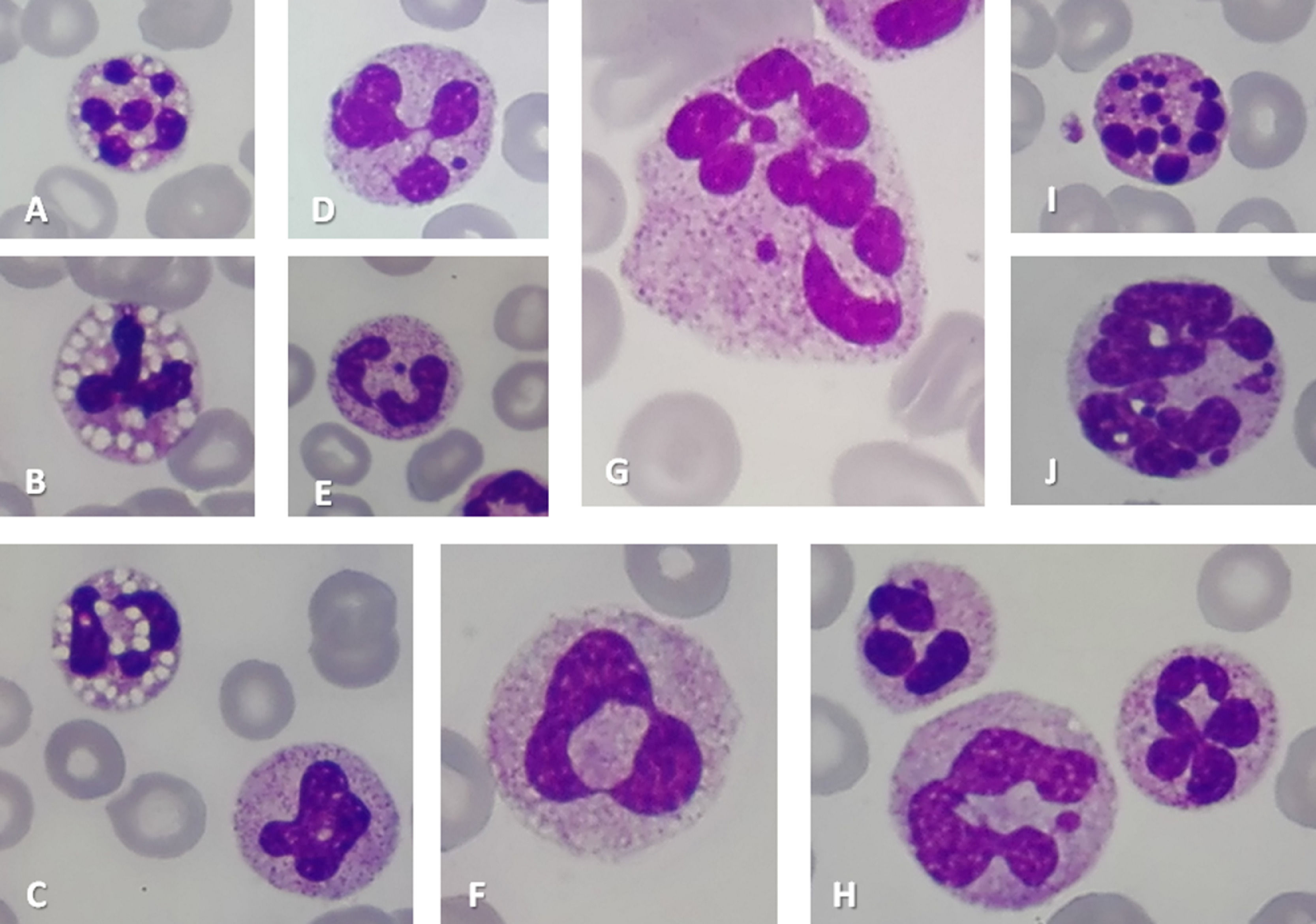
A peripheral blood film was made daily for five days and the following findings were observed (a) neutrophils with granulation, some with vacuolation or Howell-Jolly Body-like (HJBL) inclusions or ring nucleus shape, (b) macropolycytes with granulation, some with HJBL inclusions (more evident in the third day (WBC 42.60×10⁹/L, platelets count 14 × 109/L, CRP 26.6 mg/dL and procalcitonin 9.6 ng/mL)), (c) Döhle bodies in neutrophils and macropolycytes and (d) apoptotic neutrophils/macropolycytes (more evident in the third day, then decreasing) Figure 1.
 or Howell-Jolly Body-like inclusions (Images D, E) or ring nucleus shape (Images C, F), Macropolycytes (Images F, G, H, J), Macropolycytes with Howell-Jolly Body-like inclusions (Image G), Apoptotic neutrophils/macropolycytes (Images I, J) (May-Grünwald-Giemsa stain, original magnification x1000).")
Neutrophils with vacuolation (Images A, B, C) or Howell-Jolly Body-like inclusions (Images D, E) or ring nucleus shape (Images C, F), Macropolycytes (Images F, G, H, J), Macropolycytes with Howell-Jolly Body-like inclusions (Image G), Apoptotic neutrophils/macropolycytes (Images I, J) (May-Grünwald-Giemsa stain, original magnification x1000).
Serratia marcescens was isolated both from the blood culture and a vascular catheter.
Macropolycytes are polymorphonuclear leucocytes with a diameter greater than 14 microns.1 They are tetraploid and therefore represent a missed cell division.2 Macropolycytes can be found in pernicious anemia,1,3 myelodysplastic syndromes,2,4,5 therapy with granulocyte colony-stimulating factor (G-CSF)2,4,6,7 HIV infection,2,7 myeloproliferative neoplasms and megaloblastic anemia, infections2 or as described in our case report, sepsis.1,3 Very occasional macropolycytes can be seen in the blood films of healthy individuals.2,5
HJBL inclusions are detached pycnotic nuclear fragments which can be associated with immunosuppressive therapy or chemotherapy,8 ganciclovir9 and acquired immunodeficiency syndrome.4 Döhle bodies are essentially found in infectious and inflammatory conditions. Neutrophils with vacuolation are usually seen in infection and sepsis. Neutrophils with ring nucleus shape can be associated with myelodysplastic syndromes, megaloblastic anemia, infectious or autoimmune diseases10 and sepsis.
Apoptosis is most frequently associated with infection.11 However, it may also be associated with heat shock, hypoxia, cytotoxic drugs, steroids, viral infections, and radiation.12
In this case report we describe a set of characteristic hematological findings seen in sepsis, which together reinforce this diagnosis. However, if the sepsis is cured and these findings persist, a different disease may exist.


