

Sickle cell disease (SCD) is the most important hemoglobinopathy worldwide. The treatment often requires phenotype-matched red blood cell (RBC) transfusions, but alloimmunization to non-ABO antigens may occur in a part of the SCD patients. The genotyping has been used for RBC antigen prediction, reducing the possibility of the alloimmunization.
Objective and MethodIn this study we performed the genotyping for the Kell and RHCE blood groups in samples from 77 phenotyped Brazilian SCD patients, whose alloimmunization profiles were also assessed.
ResultsDiscrepancies between genotyping and phenotyping for the RHCE and Kell blood groups systems were observed in 22.07% (17/77) of the SCD patients. We found C/c and E/e discrepancies in 11.68% and 9.09% of patients, respectively; one SCD patient (1.3%) presented a discrepancy in the Kell group. Two SCD patients with discrepancies between genotype and phenotype were alloimmunized. In total, twenty-eight patients (36.4%) developed alloantibodies, of which 55.17% were directed against antigens in the Rh system, 8.62% were directed against antigens in the Kell system and 36.20%, against other groups. Finally, the frequency of discrepancies is significantly higher in non-alloimmunized patients (30.61%), compared to alloimmunized patients (7.14%) (p = 0.0217).
ConclusionIn part, the alloimmunization of the SCD patients may have been triggered by these discrepancies, indicating that the integration of serological and molecular tests in the immunohematology routine could help to increase the transfusion safety. However, the higher number of alloimmunized patients without discrepancies showed that reasons other than the discrepancies appear to have influenced more strongly the alloimmunization in the SCD patients in this study.
Considering the frequency and social impact, the sickle cell disease (SCD) is the most important hemoglobinopathy worldwide, being recently recognized as a global public health problem by the World Health Organization (WHO). This disease is characterized by a single amino acid substitution at the sixth residue of the beta (β)-globin subunit (p.Glu6Val), resulting in the production of the Hemoglobin S (HbS).1 The presence of abnormal HbS drives the pathophysiology of SCD, since the hypoxia‐induced intracellular polymerization of HbS leads to distorted cell morphology (sickling), hemolysis, altered blood viscosity, occlusion of blood flow at the level of capillaries and postcapillary venules. This vaso-occlusion leads to end-organ ischemia-reperfusion injury and infarction. In addition, the vaso-occlusive events and the intravascular hemolysis promote inflammation and redox instability, leading to the progressive small- and large-vessel vasculopathy.2 Since the polymerization of HbS is the factor that initiates the pathophysiology of the disease, the therapy is based on the reduction of HbS concentration. Currently, the use of hydroxyurea, L-glutamine and transfusions are indicated for the treatment of SCD patients.3
The transfusion with sickle negative erythrocytes is the most important treatment for SCD, able to reduce the concentration of circulating HbS, mainly for management of acute conditions and prevention of complications associated with SCD. On the other hand, the development of alloantibodies to non-ABO antigens, such as in the Kell and RHCE blood groups, is a serious hazard. Up to 50% of transfused patients with SCD may form red blood cell (RBC) alloantibodies, causing difficulties in subsequent transfusions or pregnancies and may be deadly.3,4 In this context, phenotype-matched RBCs for the most immunogenic antigens is the strategy used to avoid the alloimmunization. However, despite the phenotype-matched blood transfusions, the alloimmunization rates reach approximately 15%, in part due to the presence of variant RBC antigen alleles. So, the genotyping has been used for the RBC antigen prediction, increasing the availability of blood typed for clinically relevant blood group antigens, as the variant antigen gene alleles are often detected by this technique.5
Based on this gap in the care of SCD patients, we performed the genotyping for the Kell and RHCE blood groups in samples of these patients treated at the Base Hospital of the Federal District of Brazil. In addition, we observed the discrepancies between the genotype and phenotype and the alloimmunization profile, to better understand the impact of the matched blood transfusion based only on the serological phenotyping of RBCs for the treatment of SCD patients.
MethodsEthics considerationsThe study was approved by the Research Ethics Committee of the Health Secretariat of the Federal District, Brazil (CEP/SES/DF, protocol N° 63463316.7.0000.5553) and the informed written consent was obtained from each and all SCD patients (or a legal guardian in case of disability).
Subjects and sample collectionThe SCD patients included in this study were submitted to a transfusion protocol for SCD at the Base Hospital of the Federal District of Brazil. In summary, these patients were submitted to therapeutic transfusion and/or prophylactic transfusion. Red blood cell concentrates must respect the following patient antigens: ABO, Rh (CDE) Kell (K), Duffy (Fya, Fyb) Kidd (Jka, Jkb) and Ss. In situations in which the phenotype cannot be respected (due to insufficient stock, emergency transfusion or any other reason), the transfusion agency may authorize the transfusion with exposure to an incompatible antigen, considering the following order of immunogenicity: > RhC > RhE > Rhce > K > Jka > Fya > Jkb > Fyb > S > s. For this study, approximately 5mL of whole blood was obtained from 77 SCD patients in sterile vacutainers with EDTA (Vacuette, Greiner Bio-one, Kremsmuster, Austria). The samples were preserved under refrigeration (4 to 8°C) until the DNA extraction was performed.
DNA extractionThe genomic DNA was extracted from the peripheral blood using the Maxwell® 16 Blood DNA Purification Kit (Promega, Wisconsin, USA), according to the manufacturer's instructions. The concentration and quality of the DNA were analyzed by the optical density in the spectrophotometer NanoDrop Lite (Thermo Scientific, California USA).
RHCE genotypingFor the RHCE*C/c genotyping, PCR-SSP primer sets were used (Table 1). These sets generated PCR products of 356 bp for the RHCE*C allele and 177 bp for the RHCE*c allele. The PCR-SSP reaction was carried out in 50μl of the total volume, with 5 µL of extracted DNA (100 to 300ng/µL), 2µL of each primer (2.5 pMol), 3µL of dNTP (1.25 mM) (Invitrogen, California, USA), 5 µL of 10× buffer, 1.5µL of MgCl2, 0.5µL of (5 U/μL) Taq DNA polymerase (Invitrogen, California, USA) and 29.5µL of nuclease free water. The amplification program used was as follows: 1 cycle at 95°C for 15 minutes; 30 cycles of 94°C for 1 minute, annealing at 65°C for 1 minute and extension at 72°C for 3 minutes and 30 seconds and a final extension of 72°C for 10 minutes and subsequent maintenance at 4°C. The amplicons were visualized on a 2% agarose gel stained with GelRed (Biotium, California, USA).
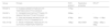
Primers and restriction enzymes.
| Group | Primers | PCR product | Restriction enzymes | RFLP a |
|---|---|---|---|---|
| RHCE*C | 5’-TCGGCCAAGATCTGACCG-3′5´-TGATGACCACCTTCCCAGG-3’ | 177bp | - | - |
| RHCE*c | 5’- CAGGGCCACCACCATTTGAA-3’5’-GAACATGCCACTTCACTCCAG-3’ | 356bp | - | - |
| RHCE*E/e | 5’- GGCAACAGAGCAAGAGTCCA-3’5’- CTGATCTTCCTTTGGGGGTG-3’ | 474bp | Mnl I | 155bp + 118bp |
| KEL*01/02 | 5’- AAGCTTGGAGGCTGGCGCAT-3’5’- CCTCACCTGGATGACTGGTG-3’ | 156bp | Mva16691 | 156bp + 98bp + 58bp |
The RHCE*E/e genotyping was performed by the PCR-RFLP assay. For this, a set of primers that generates PCR products of 474 bp was used (Table 1). The PCR reaction was carried out in 50μL of total volume, with 5 µL of extracted DNA (100 to 300ng/µL), 6 µL of each primer (2.5 pMol), 6 µL of dNTP (1.25 mM) (Invitrogen, California, USA), 5 µL of 10× buffer, 1.5µL of MgCl2, 0.5µL of (5 U/μL) Taq DNA polymerase (Invitrogen, California, USA) and 20 µL of nuclease free water. The amplification program used was as follows: 1 cycle at 95°C for 8 minutes; 35 cycles of 96°C for 1 minute, annealing at 61°C for 30 seconds and extension at 72°C for 2 minutes, and; a final extension of 72°C for 10 minutes and subsequent maintenance at 4°C. After the amplicon visualization, the PCR product was incubated with Mnl I restriction enzyme (Invitrogen, California, USA) at 37°C for 30 minutes, according to the manufacturer's recommendation. The products of the reactions were visualized on a 2% agarose gel stained with GelRed (Biotium, California, USA).
Kell genotypingThe KEL*01/KEL*02 determination was performed by PCR-RFLP assay. For this, a set of primers that generates PCR products of 156 bp was used (Table 1). The PCR reaction was carried out in 25μL of total volume, with 2µL of extracted DNA (100 to 300ng/µL), 1µL of each primer (2.5 pMol), 2µL of dNTP (1.25 mM) (Invitrogen, California, USA), 2.5µL of 10× buffer, 0.75µL of MgCl2, 0.3µL of (5 U/μL) Taq DNA polymerase (Invitrogen, California, USA) and 10.45µL of nuclease free water. The amplification program used was as follows: 1 cycle at 94°C for 2 minutes; 35 cycles of 94°C for 40 seconds, annealing at 61.5°C for 50 seconds and extension at 72°C for 1 minute and a final extension of 72°C for 10 minutes and subsequent maintenance at 4°C. After the amplicon visualization, the PCR product was incubated with the Mva 12691 restriction enzyme (Invitrogen, California, USA) at 37°C for 30 minutes, according to the manufacturer's recommendation. The products of the reactions were visualized on a 2% agarose gel stained with GelRed (Biotium, California, USA).
Serological phenotyping and alloimmunization dataThe serological phenotyping was performed as follows: I) for the Rh and Kell systems, Bio-Rad Rh/K Phenotyping cards were used (Bio-Rad, California, USA); II) for the phenotyping of Kpa, Kpb, Jka, Jkb, P1, Lea, Leb, Lua, Lub, M, N, S, s and Fya and Fyb antisera and Grifols DG Gel cards were used (Grifols, Barcelona, Spain). In addition, the alloimmunization profile was obtained using the ID-DiaPanel and the ID-DiaPanel-P for carrying out enzymatic procedures (Bio-Rad, California, USA). Data from the phenotyping and alloimmunization were inputted in the SISTHEMO - transfusion module. At the moment of blood collection, the information about the serological phenotyping and alloimmunization were obtained from this module, which is used by the healthcare system of the Federal District of Brazil.
Statistical analysisThe genotype and phenotype data were compared and the percentage of discrepancies were calculated. The comparison of alloimmunization rates between males and females and between different age ranges were performed by the Fisher's exact test (GraphPad Prism 7) and the p-value < 0.05 was considered as significant. The Fisher's exact test was also used to compare the frequency of discrepancies in alloimmunized and non-alloimmunized groups.
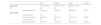
ResultsA total of 77 SCD patients were included in the study, 39 females (34.25 ± 11.90 years old) and 38 males (31.68 ± 8.98 years old). The mean age was 32.98 ± 10.57 for all patients. Twenty-eight patients (36.4%) developed alloantibodies and 49 (63.6%) did not. Although the number of alloimmunized female SCD patients (46.2%) was higher than the number of alloimmunized male SCD patients (26.3%), our data indicated that there was no significant association between gender and the rate of alloimmunization (Table 2).
Alloimmunization profile in patients with sickle cell disease according to the age and gender.
| Male | Female | Total | p-value | ||
|---|---|---|---|---|---|
| Age (±sd) | 31.68 (±8.98) | 34.25 (±11.90) | 32.98 (±10.57) | ||
| N | 38 | 39 | 77 | — | |
| Alloimmunized (n) | 10(26.3%) | 18(46.2%) | 28(36.4%) | 0.0977 | |
| Age range | |||||
| ≤ 29 years old | 18 | 17 | 35 | — | |
| Alloimmunized (n) | 4(22.2%) | 7(41.2%) | 11a(31.4%) | 0.2890 | |
| ≥ 30 years old | 20 | 22 | 42 | — | |
| Alloimmunized (n) | 6(30.0%) | 11(50.0%) | 17a(40.5%) | 0.2225 |
Considering that older SCD patients possibly have had a higher number of transfusions and, consequently, a higher risk of alloimmunization, the SCD patients evaluated were grouped into patients ≤ 29 years old and those ≥ 30 years old and the percentages of alloimmunization were observed. Although the number of alloimmunized SCD patients was higher in the ≥ 30-year-old group, no significant difference in the alloimmunization rate was observed between the groups (Table 2).
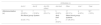
From the reported alloantibodies, 55.17% were directed against antigens in the Rh system and 8.62% were directed against antigens in the Kell system. Alloantibodies against other groups were also observed (36.20%). Considering the Rh system, the alloantibody against C had the highest frequency (20.69%), followed by the Anti-E (12.06%). Alloantibodies against other Rh antigens were observed in 22.41% and alloantibodies against the Kell group were observed in 8.62% of the SCD patients (Table 3).
Alloimmunization profile in patients with sickle cell disease according to blood groups.
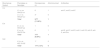
The disagreements between serological phenotyping and genotyping were observed for the RHCE and Kell blood group systems (Table 4). A total of 17 SCD patients (22.07%) presented discrepancies. Of these, 9 presented C/c discrepancies (11.68%) and 7 presented E/e discrepancies (9.09%). Only one SCD patient (1.3%) presented the discrepancy for the Kell group. Two SCD patients who presented discrepancies were alloimmunized. One patient phenotyped as C+c+ and genotyped as RHCE*c/c developed alloantibodies against C, D and E. Another patient phenotyped as E-e+ and genotyped as RHCE*E/e developed anti- E, anti-C, anti-D, anti-V, anti-VS, anti-G, anti-S and anti-Dia alloantibodies.
Discrepancies between genotype and phenotype.
| Blood group System | Phenotype vs. Genotype | Discrepancies (n) | Alloimmunized | Antibodies |
|---|---|---|---|---|
| C/c | ||||
| C+ c+ vs. RHCE*c/c | 6 | 1 | anti-C, anti-D, anti-E | |
| C- c+ vs. RHCE*C/c | 3* | 0 | ||
| Total | 9/77 (11.68%) | 1 | ||
| E/e | ||||
| E+ e+ vs. RHCE*e/e | 1 | 0 | ||
| E- e+ vs. RHCE*E/e | 6 | 1 | anti-E, anti-C, anti-D, anti-V, anti-VS, anti-G, anti-S, anti-Dia | |
| Total | 7/77 (9.09%) | 1 | ||
| Kell | ||||
| K- k+ vs. KEL*01/02 | 1* | 0 | ||
| Total | 1/77 (1.3%) | 0 |
Finally, the Fisher's exact test (Table 5) demonstrated that the frequency of discrepancies is significantly higher in non-alloimmunized SCD patients (15/49 or 30.61%), compared to alloimmunized SCD patients (2/28 or 7.14%) (p = 0.0217).
Discrepancies in alloimmunized and non-alloimmunized groups.
| With discrepancies | Without discrepancies | Total | p-value* | |
|---|---|---|---|---|
| Alloimmunized | 2 (7.14%) | 26 (92.86%) | 28 (100%) | 0.0217 |
| Non-alloimmunized | 15 (30.61%) | 34 (69.39%) | 49 (100%) | |
| Total | 17 | 60 | 77 |
While transfusions are effective in preventing morbidity in patients with SCD, the alloimmunization against foreign RBC antigens is a major challenge in a transfusion. Patients with SCD with multiple transfusions notoriously produce more alloantibodies to RBC antigens than any other patient population for reasons that are not completely understood. The RBC phenotypic disparity conditioned by ethnic differences between donors and patients partially explain this higher rate of alloimmunization, which especially affects C, E and K antigens.6 Here, we observed that 36.4% of the SCD patients developed alloantibodies. In Brazil, the incidence of alloimmunization in patients with SCD ranges from 10 to 60%.7 Worldwide, variable alloimmunization rates were also reported in patients with SCD. In the USA, previous studies revealed the alloimmunization rates of SCD patients variating from 7.07% to 58%.8,9 In the UK, alloimmunization rates ranged from 17.65 to 76.19%. In the Middle East, alloimmunization rates ranged from 13.71% in Saudi Arabia to 65.45% In Kuwait.8 In some regions, the incidence of alloimmunization is lower. In sub-Saharan Africa (SSA), the overall proportion of alloimmunization was 7.4%.10 Among Palestinian SCD patients, the frequency of RBC alloimmunization was 7.76%.11 Moreover, immune antibodies occurred in 2.6% of the Jamaican SCD patients.12 These low proportions of alloimmunization may be related to the close racial background between donors and SCD patients. In addition, factors, such as the heterogeneity of efficacy of the alloimmunization prophylaxis protocols, the occurrence of occasional transfusions in centers without an antigen-matching transfusion policy, the rate of RH variants in each studied population and the absence of matching units in the emergency scenario, may explain the differences in alloimmunization rates among the studies.7,13
In this study, 55.17% of the reported alloantibodies were against Rh antigens and 8.62%, against Kell antigens, despite the primary matching for C/c, E/e and K antigens. Among the alloantibodies against Rh antigens, 20.69% was the alloantibody against C and 12.06%, against E. The alloantibodies against the Rh and Kell systems comprise the majority of the antibodies detected in SCD patients. The relatively high immunogenicity of the Rh and Kell antigens and the antigenic discrepancy between the donor and the recipient are considered the most important contributing factors. Furthermore, the higher rate of alloimmunization against Rh (D, C/c and E/e) antigens in SCD patients, even with serologic matching, may be explained by the presence of an increased prevalence of the RHD and RHCE variants in this population.4
Although the use of serological phenotyping to select phenotype-matched RBCs has become a standard of care for reducing the alloimmunization incidence, the genotyping provides additional information to avoid the incompatibility of RBC antigens between the donor and the recipient.6 In fact, we observed discrepancies between serological phenotyping and genotyping for the RHCE and Kell blood group systems in 22.07% of the SCD patients. Considering the alloimmunized SCD patients, discrepancies were found in 7.14% (2/28). In Brazil, a higher percentage of discrepancies between the previous phenotype and genotype-derived phenotype was found in alloimmunized SCD patients.14,15 The high cost of genotyping prevents its use in the routine of our blood bank service, leaving only serological phenotyping of RBCs prior to the transfusion. However, this prophylaxis protocol may fail, leading to the alloimmunization.
As related, we observed the alloimmunization in two SCD patients with discrepancies between genotyping and serological phenotyping for the RHCE blood group. A patient phenotyped as C+c+ and genotyped as RHCE*c/c developed anti-C alloantibodies. This observation suggests the presence of RHCE variant alleles. Furthermore, approximately 20% of the SCD patients with C+ red cells express variant C without a conventional C antigen. This variant C is encoded by a hybrid RHD*DIIIa-CE(4-7)-D gene that is actually located in the RHD locus that does not produce the D antigen, but a variant C antigen, leading to a C+ serologic phenotype. When repeatedly exposed to conventional C+ red cells, some patients with this RHD hybrid gene develop the anti-C against the conventional C+ protein.16 Additionally, due to this hybrid allele, SCD patients phenotyped as C+ are better served with C- transfusion to avoid the risk for allo-anti-C.17 Another patient phenotyped as E-e+ and genotyped as RHCE*E/e developed anti-E alloantibodies. Unexplained Rh antibodies in patients with SCD were occasionally observed in previous studies, including anti-E in both E+ and E− patients, despite the Rh matching.8 Another hypothesis is the presence of a rare Rh Ew variant in this SCD patient. According to some reports in the literature, anti-E alloantibody was found in an Ew positive patient, after receiving E positive red blood cells.18,19 On the other hand, the DNA sequencing is necessary to confirm the presence of RHCE variants in these patients. Unfortunately, the RHCE sequencing was not performed due to the considerable financial investment it requires, as well as the need for a specialized laboratory with expertise in blood group genetics to interpret the results. Further studies will investigate the alloimmunization by the RHCE sequencing in our population.
The discrepancies between serological and molecular analysis for the Kell antigen were observed less frequently, when compared to the Rh antigens. Only one SCD patient (1.3%) presented the discrepancy for the Kell group. As in our study, the KEL allele discrepancies were found in 5% of the alloimmunized thalassemia patients from Iran. Interestingly, most of the discrepancies were related to the K-k+ phenotype status and the KEL*01/KEL*02 genotype.20 The occurrence of these cases may be due to new alleles in the Kell blood group locus that lead to the variant Kell phenotype.
Finally, both gender and age were not statistically significantly associated with the risk for the alloimmunization in our study, despite the fact that the alloimmunization was higher in women and in the group over 30 years old. In general, the alloimmunization prevalence rates are higher in females than in males.21,22 On the other hand, comparable rates were reported in adults with SCD,23 though the history of pregnancy may also be associated with an increased risk for alloimmunization and impact these data. Regarding the transfusion recipient age, the alloimmunization prevalence increases with age both in general and SCD patients,20–24 with the number of RBC transfusions being a significant risk factor.25 Unfortunately, it was not possible to obtain reliable data on the number of transfusions throughout life for the SCD patients in our study, which prevents us from concluding whether the number of transfusions actually influences the higher rate of alloimmunization found in older patients.
Despite the discrepancies between serological phenotyping and genotyping possibly triggering an alloimmunization process after the incompatible transfusion, most alloimmunized SCD patients did not have discrepancies in this study. Therefore, the alloimmunization process may be triggered by different factors than discrepancies. We believe that a deviation from the main transfusion protocol (such as in cases of emergency transfusion or low compatible blood supplies) exerts a strong influence for the alloimmunization in our patients. Frequently, SCD patients are assisted at different hospitals and the absence of centralized records to ensure the availability of transfusion history and immunohematology information may also lead to the alloimmunization. To prevent this outcome, transfusion protocols must be created. At our institution, a guideline to guarantee transfusion safety in relation to the compatibility of the Rh and Kell antigens was introduced. The genotype results for all patients were considered for transfusion purposes. Furthermore, as a prophylaxis protocol for all SCD patients, the alloantibodies profile is considered to define transfusional units and to perform phenotype-matched blood transfusions.
ConclusionIn conclusion, we investigated the disagreements between serological phenotyping and genotyping for the RHCE and Kell blood groups systems. Furthermore, we observed the presence of alloantibodies that, in part, could be originated by these discrepancies. However, other reasons (such as transfusion protocol deviation) appear to exert a higher influence on the alloimmunization in the studied population. Despite the use of antigen-matching by serology reducing the risk of alloimmunization, the RHCE and Kell genotyping of SCD patients and donors can prevent the unwanted alloimmunization and should be encouraged. Considering that the alloimmunization is an important complication for SCD patients, further studies are needed to clarify the other risk factors that lead to the alloimmunization. Thereby, specific transfusion protocols to prevent this outcome could be adopted.


