
Treatment-free remission (TFR) is successful in half of the patients with chronic myeloid leukemia who discontinue Imatinib (IM) after sustained molecular response.
MethodsIn a prospective trial, we used pioglitazone for 3 months before stopping IM in 30 patients. Percentages of peripheral blood lymphocyte subsets were assessed before and after treatment. The relation of these data with duration of IM treatment and TRF were examined.
ResultsThe median time of IM treatment was 117.6 months. After discontinuation, 11 patients had molecular recurrence after 5.2 months (2.4 – 30). The observation time for those remaining in TFR was 46 (26 – 56) months. The independent factors for the maintenance of TFR were the duration of IM treatment and the percentage of double-positive T cells at IM stop.
ConclusionA longer treatment with imatinib was associated with a longer TFR after discontinuation. Pioglitazone could act as an immunomodulator, increasing DP T cells which may contribute to prevent relapse.
In recent years, several attempts have been made to stop tyrosine kinase inhibitor (TKI) treatment in chronic myeloid leukemia1–6 after patients have achieved a sustained deep molecular response. Besides, the immune system plays a role in maintaining TFR after TKI discontinuation. It has been described that NK and cytotoxic T lymphocytes are involved in this process.6,7 Ilander et al.8 and Rea9 have shown in the IMMUNOSTIM trial, that patients maintaining treatment-free remission (TRF) presented more natural killer cells of the cytotoxic CD56dim subset.8 No association was found between patients’ relapse and the number of CD3+/CD4+/CD8+ T (DP T) cells, the CD4/CD8 ratio, or Tregs. A sub-analysis from the EURO-SKI study has demonstrated that low CD86+ plasmacytoid dendritic cells, which mediate immune tolerance, might predict TFR in patients discontinuing TKI treatment.2
Recently, we have conducted a prospective controlled phase II imatinib (IM) discontinuation study (EDI-PIO trial - in Portuguese: Estudo de Descontinuação de Imatinibe após Pioglitazona) in patients with CML treated at our Institution.4 In this study, we administered pioglitazone for 3 months before stopping imatinib as it has been shown that this drug, that is a proliferator‐activated receptor (PPAR)‐γ agonist used for diabetes treatment is able to pull the CML leukemic stem cells (LSC) out of quiescence and sensitizing them to imatinib in pre-clinical models.10
In the present work we evaluated the immunological profile of CML patients included in this trial and compared its influence on long-lasting TFR of the patients with the time of IM treatment. We analyzed the peripheral lymphocyte subsets before starting pioglitazone and at the stop of both, IM and pioglitazone, and the total time of IM treatment.
Patients and methodsStudy designEDI-PIO was a prospective, open-label, phase 1/2, single-center, single-arm pilot study of IM discontinuation, in adult patients with CML in chronic phase with stable deep molecular response.4 The protocol was approved by the Institutional Review Board of the University of Campinas and all patients gave written informed consent. The study is registered at ClinicalTrials.gov, number NCT02852486.
We included patients followed in our Institution between 1998 and 2015, with CML in chronic phase, imatinib treatment longer than 3 years, deep stable 4.5 log molecular response (BCR-ABL::ABL1. ratio ≤ 0.0032%), confirmed by 4 RQ-PCR tests in the last 2 years (2 tests within the last 6 months). The imatinib dose at discontinuation was 400 mg/day. Only one patient was receiving 300 mg/day. We excluded patients with CML in accelerated phase (AP) or blast crisis (BC), Philadelphia positive (Ph+) acute lymphoid leukemia and CML patients presenting BCR-ABL mutations related to resistance. Patients were treated with pioglitazone (EMS S/A, Brazil) 30 mg/day for 3 months before imatinib discontinuation. BCR-ABL levels were measured by real-time quantitative PCR (RQ-PCR) monthly after imatinib discontinuation during the first 12 months, every two months in the second year, and then every 3 months during the subsequent follow-up. Results were reported by using the BCR-ABL International Scale.11 Molecular relapse was defined as any single sample with a value of > 0.1% (loss of MMR) or two consecutive positive samples with a value > 0.01% (MR4.0), with 4 week-interval. Imatinib was re-introduced at the same dose used before discontinuation if molecular relapse occurred.
ImmunophenotypingAn 8-color panel of monoclonal antibodies was used to examine the several lymphocyte subsets in peripheral blood before start of pioglitazone and at stop of imatinib + pioglitazone treatment. Antibodies against CD3, CD4, CD8, CD16, CD19, CD27, CD45, CD45RA, CD45RO, CD56, CD57, CD62L, TCRαβ and TCRγ were used in an 8-color platform. The acquisition was made in a FACSCanto II equipment, analyzing 100.000 events. We assessed the percentages of B lymphocytes, as well as T(CD4+), T(CD8+), T(CD4+CD8+), T(CD4-CD8-), TCRαβ, TCRγδ, Tnaive, Tmemory, NK and NK-T cells.12
Statistical analysisDescriptive analysis was performed using non-parametric tests. Differences among groups and relations between lymphocyte subsets before and after pioglitazone were analyzed by the Mann–Whitney test. The influence of clinical data at diagnosis, TKI treatment duration and lymphocyte subsets at the start of pioglitazone and at the stop of IM and pioglitazone was analyzed by univariate and multivariate Cox models using stepwise analysis (backward conditional method). Variables entering the multivariate model were selected by p = 0.05 for inclusion and p = 0.10 for exclusion.
ResultsBetween June 2016 and January 2019, 32 patients from EDI-PIO trial received pioglitazone and 31 discontinued therapy. Results concerning peripheral blood lymphocyte subsets could be retrieved in 30 cases. The demographic features of the patients are shown in Table 1. The majority of the cases had BCR::ABL1 transcript b3a2, and had a low risk by Sokal score. Six patients had previously received Interferon–alpha for a median of 11 months (1–30) before starting IM. All patients had received pioglitazone for three months before stopping IM. The median time of TKI treatment was 117.6 months (40 - 191). In 23 (74%) cases, a cytogenetic remission was achieved within the first year of treatment. A molecular remission MR4.5 was achieved in a median of 31 months (5 – 83). After IM discontinuation, 11 patients had a molecular recurrence in a median time of 5.1 months (2.4 – 29.5) and Imatinib was reintroduced. The 19 patients remaining in sustained TFR had a median observation period of 55 months (35 – 69).
Demographic features of the CML patients (n = 30).
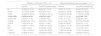
The percentages of the peripheral lymphocyte subsets examined before the start of pioglitazone and at IM stop are shown in Table 2. There was no significant difference for patients remaining in TFR or recurring for any subset analyzed at the start of pioglitazone. At IM+pioglitazone discontinuation, the only lymphocyte subset that increased in patients that maintained TFR but not in patients with molecular relapse was the percentage of double positive CD4+ / TCD8+ T cells (DP T) (p = 0.002).
Distribution of the peripheral lymphocyte subsets (median and range) at TKI discontinuation in percentage among all peripheral blood lymphocytes.
In the univariate Cox model, the following variables were related to the time of TFR: duration of IM treatment B= −0.001, exp(B) 0.999 (CI 0.998 – 1.00), p = 0.005; time to achieve MR4.5 B = 0.00, exp(B) 1.00 (CI 1.00 – 1.00), p = 0.027; DP T cells at IM stop B = −0.485 exp(B) 0.615 (CI 0.408 – 0.929), p = 0.021 and T cells gamma delta (γδ) at IM stop B = 0.285 exp(B) 1.329 (CI 1.029 – 1.717), p = 0.029. Type of transcript, Sokal score, previous use of interferon alpha, and all the peripheral lymphocyte subsets at start of pioglitazone had no significant influence in TFR time in the univariate Cox model.
The best multivariate model predicting maintained TFR was found for the duration of IM treatment (B = −0.001 p = 0.064) and DP T cells at IM stop (B = −0.339 p = 0.095).
DiscussionIn the present study, analyzing data concerning duration and response of TKI treatment and the peripheral lymphocyte subpopulations in patients with CML treated for a long period with imatinib, we found that only the increase in the proportion of DP T cells after pioglitazone was related to the maintenance of TFR after imatinib discontinuation. Besides, γδ T lymphocytes were slightly lower in patients maintaining remission.
The rationale for using pioglitazone in the present trial was based on a previous investigation of Prost et al.10 They reported 3 patients in which improvement in molecular responses was observed after simultaneous administration of pioglitazone and imatinib. Later, the same group published preliminary findings of the ACTIM study, where 24 patients received pioglitazone and imatinib for one year. This group of patients had a higher rate of molecular response (MR4.0) compared to a historic control.13 Besides, a trial conducted in India combined pioglitazone with imatinib to improve responses in CML patients with sub-optimal responses with TKI.14 A one log reduction after pioglitazone was observed in 89% of patients. In both studies, the peripheral lymphocyte subsets after pioglitazone were not analyzed.
Other discontinuation trials analyzed the immunological profile of CML patients by flow cytometry. Recently, Luo et al.15 showed that NK cells were increased at baseline in CML patients who did not relapse after stopping TKI compared to relapsing patients, but they did not analyze DP T cells. Irani et al. also reported that patients who had a successful TFR had a higher proportion of NK cells at TKI discontinuation, but they did not evaluate the role of DP T cells.16
The role of the immune system in the outcome of CML has been studied but there are few data on which are the most important cell types.8,16,17 At diagnosis, patients with CML present impairment of the innate immune response, including NK cells, dendritic cells as well as cytotoxic T cells.6 Treatment with IFN-α restores NK number and function is 3–6 months. But also TKI treatment increases the number of NK cells compared to diagnosis.13 This feature was not examined in the present study. Besides, CD8+ cytotoxic cells increase and have their function restored after TKIs and are associated with a better response to therapy.6 Double-positive T cells do exist in normal individuals.18 Their function is not completely understood, but they have a high cytotoxic potential. They are increased in the elderly, in neoplasms such as melanoma and in auto-immune diseases.18-20 So, the DP T cells could have a cytotoxic or immunomodulating effect that could contribute to TFR maintenance. So, the DP T cells could have a cytotoxic or immunomodulating effect that could contribute to TFR maintenance but, to the best of our knowledge, this has not yet been examined in CML. On the other hand, Seguro et al. demonstrated in an imatinib discontinuation trial that the NK cell proportion at baseline was lower in CML patients in MR4.0 who relapsed after discontinuation. However, the authors did not find a relationship between NK proportion and relapse when patients discontinued therapy in MR4.5,6 similar to our findings.
In the present study, the rationale for using pioglitazone was to alter the cell cycling of the leukemic stem cells, to increase imatinib effect in this population before discontinuation. One unexpected finding was the higher number of DP T cells after pioglitazone exposure in the non-relapsing patients. Pioglitazone causes several metabolic changes in CML cells, such as mitochondria fatty acid overload, dysfunction, and oxidative damage, as demonstrated in a previous study of our group.21 Fu et al. showed that pioglitazone restores IFN-γ production in tumor-infiltrating invariant natural killer T cells from both human patients and mouse melanoma models.22 In a mouse model of type 2 diabetes, pioglitazone induced an increase of T lymphocytes and CD3+IL-4 production in immigrant spleen lymphocytes.23
Our findings suggest that in some patients, pioglitazone is able to recruit double-positive T cells, and perhaps stimulate immunity to an anti-leukemic effect. In the multivariate analysis the results were not significant, probably due to the small sample size, one limitation of our study.
Recurrence was observed only in the first 3 years after discontinuation, while in patients who remained in TFR the observation period was always > 3 years (median 55 months). The optimal time of TKI treatment is still controversial, and our results show that a long treatment time was associated with a longer time of TFR. Among our patients, no recurrence was observed in those treated with TKI for longer than 8 years.
In conclusion, a longer time of treatment with imatinib is necessary to maintain patients with CML in TFR. Pioglitazone could not only increase the sensitivity of leukemic stem cells to imatinib, but may also induce an immunological effect that could help to prevent relapse. This hypothesis should be examined in further experiments.
I.L.M. and K.M have grants from the National Research Council (CNPq) (305110/2018-7 and 309910/2018-8 respectively). A.B.P.L. was supported by Coordenação de Aperfeiçoamento de Pessoal de Nível Superior (CAPES) grant (88882.434900/2019-01).


