

To evaluate factors predictive for relapse in a cohort of adult patients with acute promyelocytic leukemia monitored by molecular methods during consolidation and during at least one month of maintenance therapy.
MethodsThe charts and laboratory data of 65 adult patients with acute promyelocytic leukemia treated according to the International Consortium on Acute Promyelocytic Leukemia 2006 protocol were reviewed. The identification of the promyelocytic leukemia-retinoic acid receptor-alpha gene rearrangement at diagnosis, post-induction, post-consolidation and during maintenance treatment was performed by qualitative and quantitative reverse transcription polymerase chain reaction.
ResultsEighty-nine patients were diagnosed with acute promyelocytic leukemia over a seven-year period and of these 65 were eligible for treatment with the protocol. Among the 45 patients who received consolidation and maintenance treatment, six (13%) relapsed, three of whom presented hematologic and three presented molecular relapse. The first relapses occurred at a median of 39 months. Relapsed patients were from all risk groups (low, intermediate and high) and both morphological types (M3 and M3variant) were found. Three of these patients are alive and in molecular remission after salvage treatment. There were no statistically significant differences regarding gender, age, risk group, morphology, promyelocytic leukemia breakpoint cluster region, use of all-trans retinoic acid, development of differentiation syndrome and number of days to complete remission between the patients who relapsed and those who did not.
ConclusionOur results reinforce the importance of prolonged monitoring of acute promyelocytic leukemia patients using molecular methods to detect relapse early.
Once considered a leukemia with poor prognosis, currently by means of adequate treatment, acute promyelocytic leukemia (APL) is highly curable, with remission rates of 90% and disease-free survival at six years of over 80%.1 Despite the increase in overall survival, cases of relapse still occur and need to be promptly identified, thus allowing pre-emptive treatment in the case of molecular relapses or re-induction in cases of hematologic relapse.2
The International Consortium on Acute Promyelocytic Leukemia (IC-APL 2006) aimed to deploy a network of National and International centers to improve diagnosis, treatment, monitoring of treatment response and support therapy and as a consequence the survival of patients with APL in developing countries. The treatment was based on the APL 2005 protocol of the Programa Espanhola de Hematologia (PETHEMA) group, adapted to the reality of the participating countries (Brazil, Mexico, Chile, Uruguay) with the substitution of the anthracycline idarubicin for daunorubicin which is more affordable and is widely used in the treatment of other leukemias.3 Of the eight Brazilian centers participating in the IC-APL, the center of this study is the only representative of the northeastern region and included almost one third of the patients of the study.
The recently published results of the IC-APL3 demonstrated an improvement in early mortality rates and overall survival compared with historical controls.4 In the present study, a cohort of patients diagnosed with APL was evaluated for risk factors associated with relapse.
MethodsPatientsThis study retrospectively analyzed 89 adult patients diagnosed with APL between January 2007 and August 2014 at the Hospital of the Fundação de Hematologia e Hemoterapia de Pernambuco (Hemope), Recife, Brazil. Clinical and laboratory data were obtained from the patients’ charts. The project was approved by the Research Ethics Committee of the institution (#028/2006) and was conducted after informed consent was obtained from the patients.
Diagnosis and monitoringThe diagnosis was established by clinical, cytomorphological, immunophenotypic and molecular criteria.5 The identification of the promyelocytic leukemia-retinoic acid receptor-alpha (PML-RAR¿) gene rearrangement was performed by reverse transcription polymerase chain reaction (nested RT-PCR and/or RT-qPCR) according to the international BIOMED-1 and BIOMED-2 protocols.6,7 Samples for RT-qPCR were sent to the reference laboratory of the IC-APL study in the Faculdade de Medicina de Ribeirão Preto da Universidade de São Paulo (FMRP-USP).
Molecular tests were performed using bone marrow (BM) samples obtained at initial diagnosis, post-induction, post-consolidation (after the third course of consolidation) and during maintenance. Standard monitoring recommended tests every three and six months, respectively, during maintenance and after treatment for up to two years.
DefinitionsComplete hematologic remission (CR) and relapse were defined using conventional criteria.8 Hematologic relapse was diagnosed based on the presence of >20% blasts or abnormal promyelocytes in the bone marrow any time after hematologic remission. Molecular remission (MR) was defined as a negative RT-PCR result of bone marrow cells after the third and last cycle of consolidation.9 Molecular persistence was defined as PCR positivity in two consecutive BM samples collected within a minimal interval of 15 days after the end of consolidation therapy. Molecular relapse was defined as the reappearance of the PML-RAR¿-specific band in two consecutive BM samples collected at an interval of 15 days at any time after consolidation therapy.3,10 Arbitrarily, early relapse was defined as that occurring within two years after proven CR and late relapse beyond this period.11 Differentiation syndrome (DS) was defined according to standard criteria.12,13
TreatmentThe standard treatment protocol used was the IC-APL 2006 with patients being categorized according to the risk of relapse as described elsewhere.3,9,14,15 The treatment options of the relapsed cases included re-induction with all-trans retinoic acid (ATRA), chemotherapy and arsenic trioxide (ATO) followed by stem cell transplantation (SCT) whenever possible.2,16–18
Statistical analysisStatistical analysis was performed using the Stata 12.0 software. The t-test was used to compare the groups regarding age, days to CR, and months of follow up. Fisher's exact test was used for categorical variables (gender, risk of relapse, morphology and type of transcript). Relative risk (RR) was calculated with a 95% confidence interval (95% CI). p-Values <0.05 were considered statistically significant.
ResultsEighty-nine patients were diagnosed with APL during the period and of these, 65 were eligible for treatment using the IC-APL 2006 protocol. The median age at initial diagnosis of the relapsed patients was 37.5 years (range: 24–46 years). Hematologic remission was achieved with a median of 32 days (range: 27–56 days).
As the main purpose of this study was to evaluate factors predictive for relapse, analysis was restricted to the 45 patients who achieved remission and who were monitored during consolidation and maintenance. Therefore, 20 out of 65 patients were excluded because they died during induction or consolidation. Six patients out of 45 (13%) presented molecular (three) or hematologic (three) relapse. The first relapse among the patients occurred at a median of 39 months (range: 21–48 months) following initial treatment. Median follow-up of the six patients from diagnosis was 46.5 months (range: 28–71 months) and mean number of sequential samples analyzed during monitoring was 12 per patient (range: 5–21 months).
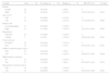
The main features of the six relapsed cases are shown in Table 1. Four patients achieved MR after salvage treatment and were subjected to autologous (auto)-SCT (Cases 1, 3, 4 and 5); two of these are alive and in MR. One patient (Case 6) was in consolidation phase of the treatment until the closing of this article.
Main features of relapsed patients from a total of 65 cases of acute promyelocytic leukemia of the Fundação Hemope.
| Case | Age (years) | Gender | Risk | Morphology | Transcript type | ATRA in induction phase (days) | Hematologic remission (days) | Relapse | Salvage treatment | Status (months) | |||
|---|---|---|---|---|---|---|---|---|---|---|---|---|---|
| Phase | Type | Place | Time (months) | ||||||||||
| 1 | 39 | M | I | C | bcr1 | 29 | 27 | PT | Late | Molecular | 35 | ATRA+CT+ATO+Auto SCT | Death (42) |
| 2 | 24 | F | I | C | bcr2 | 31 | 56 | Mn | Early | Hematologic | 24 | ATRA+ATO | Death (44) |
| 3 | 46 | M | H | C | bcr1 | 29 | 32 | PT | Late | Molecular | 48 | ATO+Auto SCT | Death (71) |
| 4 | 36 | M | L | C | bcr3 | 30 | 26 | PT | Late | Molecular | 43 | ATRA+CT+ATO+Auto SCT | Alive (64) |
| 5 | 29 | F | H | C | bcr1 | 32 | 31 | Mn | Early | Hematologic | 21 | ATRA+ATO+Auto SCT | Alive (28) |
| 6 | 40 | M | H | V | bcr1 | 27 | 44 | PT | Late | Hematologic | 47 | ATRA+ATO | Alive (49) |
M: male; F: female; L: low; I: intermediate; H: high; C: LMA-M3 classical; V: LMA-M3 variant; PT: post treatment; Mn: maintenance; ATO: arsenic trioxide; ATRA: all-trans retinoic acid; auto SCT: autologous stem cell transplantation; CT: chemotherapy.
Two patients had a second relapse, one patient (Case 2) twelve months after the first hematologic relapse while in salvage treatment waiting for auto-SCT. This patient had no human leukocyte antigen (HLA)-compatible donor and was treated with ATO without achieving a third remission. The other patient (Case 1) presented a second hematologic relapse fourteen months after the first molecular relapse. He was submitted to ATO treatment followed by auto-SCT, but relapsed 3 months later and died.
Table 2 shows the main clinical and laboratorial features of patients with or without relapse. No significant difference was observed between the two groups of patients regarding gender, age, risk of relapse, morphology, PML breakpoint, duration of ATRA treatment during induction, frequency of evolution of DS and number of days to CR.
Analysis of 45 patients with acute promyelocytic leukemia treated at Fundação Hemope considering the cumulative incidence of relapse.
| Variable | All (n=45) | No relapse (n=39) | Relapse (n=6) | RR (95% CI) | p-Value |
|---|---|---|---|---|---|
| Gender | |||||
| Female | 22 | 20 (91%) | 2 (9%) | 1 | – |
| Male | 23 | 19 (83%) | 4 (17%) | 1.91 (0.39–9.41) | 0.413 |
| Age (years) | |||||
| ≤40 | 33 | 29 (88%) | 4 (12%) | 1 | – |
| >40 | 12 | 10 (83%) | 2 (17%) | 1.37 (0.29–6.57) | 0.692 |
| Relapse risk | |||||
| Low/intermediate | 31 | 28 (90%) | 3 (10%) | 1 | – |
| High | 14 | 11 (79%) | 3 (21%) | 2.21 (0.51–9.63) | 0.283 |
| Morphology | |||||
| Classical | 43 | 37 (88%) | 5 (12%) | 1 | – |
| Variant | 3 | 2 (67%) | 1 (33%) | 2.80 (0.46–16.9) | 0.291 |
| Transcript type | |||||
| bcr1/bcr2 | 30 | 25 (83%) | 5 (17%) | 1 | – |
| bcr3 | 14 | 13 (93%) | 1 (7%) | 0.43 (0.05–3.33) | 0.391 |
| ATRA in induction phase (days) | |||||
| ≤30 | 32 | 29 (91%) | 3 (9%) | 1 | – |
| >30 | 13 | 10 (77%) | 3 (23%) | 2.46 (0.57–10.6) | 0.220 |
| Differentiation syndrome | |||||
| No | 33 | 28 (85%) | 5 (15%) | 1 | – |
| Yes | 12 | 11 (92%) | 1 (8%) | 0.55 (0.07–4.24) | 0.552 |
| Hematologic remission (days) | |||||
| ≤30 | 14 | 11 (79%) | 3 (21%) | 1 | – |
| >30 | 31 | 28 (90%) | 3 (10%) | 0.45 (0.10–1.96) | 0.283 |
RR: relative risk; CI: cumulative incidence.
The 13% incidence of relapse in this study with 45 patients from a single center is consistent with other works. Cassinat et al.19 reported a relapse rate of 14.6% among 260 patients with APL in a multicenter study in France. Moreover, Karim et al.20 in a study with 26 patients, found a 11.5% relapse rate of both molecular and of the central nervous system (CNS). In the matched-pair analyses of the PETHEMA LPA-2005 and IC-APL-2006 studies,21 relapse rates were 7.4% and 5%, respectively. No CNS relapse was detected in this series. All PETHEMA/Gruppo Italiano Malattie Ematologiche Maligne dell’Adulto (GIMEMA) risk groups were represented among relapsed patients with proportions similar to other studies.22,23 The only patient with low risk for relapse had a bcr3 PML variation which is considered of adverse prognostic factor.24
The results of this study are consistent with published data regarding age and gender of the relapsed APL patients.11,22 Both morphological types (M3 and M3v) were found among relapsed patients. In one of the three cases of hematologic relapse, there was a change in morphology from variant (M3v) to classical type (M3). Morphological changes at relapse in APL may not be a rare event, and the leukemic cells can show variable morphological features at the time of relapse, which could result in misdiagnosis as a different type of acute myeloid leukemia as pointed out by Yoshii et al.23 This author recommends a comprehensive approach to the diagnosis and appropriate treatment of relapsed APL.
The overall incidence of DS reported in the LPA series varies from 7.8% to 48%.25,26 In the current series, one case, that presented with DS during the initial treatment, suffered hematologic relapse but is alive. The European APL Group reported an increased risk of relapse independent of white blood cell count in patients developing DS.27 Thépot et al.28 suggested that the total duration of treatment with ATRA of less than 21 days during the induction phase was associated with greater rates of relapse of leukemia. In this series, all patients who relapsed were treated with ATRA for more than 21 days with an average of 30 days.27–29 No case of resistance to ATRA was identified as all treated patients achieved remission.
Current studies have shown high rates of hematologic relapse compared to molecular relapse.3,11,22 In the current series, three relapses were molecular and three hematologic. Molecular monitoring of the PML-RAR¿ transcript is useful to anticipate overt hematologic relapse10 with the IC-APL 2006 protocol recommending tests every three months for at least two years after completing treatment.3 This approach is important because the majority of the relapses in this study occurred during this period with a median of 44.5 months. In this regard, two cases of hematologic relapse in this series did not strictly follow the monitoring protocol. Although the mean number of samples analyzed per patient was higher than that reported by Cassinat et al.,19 who analyzed an average of six samples per patient (range: 1–28), there is a need for more strict monitoring.
Proposed modalities of therapy in the salvage setting for APL patients includes treatment or retreatment with ATRA, ATO, or both, cytotoxic chemotherapy, auto or allogeneic SCT and enrollment in a clinical trial.2,16–18,29 Patients treated with salvage chemotherapy alone have lower survival compared to those submitted to SCT.17 Auto-SCT should be performed when the patient achieves MR, but may increase the risk of a new relapse. Patients submitted to auto-SCT have higher rates of overall and disease-free survival than those who undergo allogeneic SCT.17 Allogeneic SCT is indicated when the patient does not achieve molecular response after consolidation. These patients seem to have more prolonged remissions and less incidence of new relapse, but a higher death rate due to graft-versus-host disease (GVHD).
The protocol for relapsed patients included ATRA and Idarubicin plus ATO followed by auto-SCT, after confirmation of the MR. Four patients did not receive anthracycline due to clinical conditions, including one patient who had a heart attack at the time the relapse was diagnosed.
The IC-APL 2006 protocol used in the present study significantly reduced early mortality and improved the overall survival of APL patients3 when compared to historical controls.4 Increased knowledge of the outcome in the subpopulation of treated patients that exhibit relapse is crucial to understanding the pathophysiology of APL and to improve survival.22 In the real-world circumstances of the healthcare delivery system, costs and availability become issues for patients and clinicians, and show the importance of single center results. This study is the first to report on molecular and hematologic relapse in a cohort of Brazilian APL patients submitted to the IC-APL 2006 protocol.
ConclusionThere were no differences in clinical-biological variables between patients with or without relapse. The data of this study also show the efficacy of the treatment protocol used for APL and the importance of prolonged monitoring using molecular methods to detect relapse earlier in order to improve survival of this subgroup of patients.
Conflicts of interestThe authors declare no conflicts of interest.
This study was supported by the American Society of Hematology and Fundação de Amparo à Ciência e Tecnologia do Estado de Pernambuco–FACEPE. The authors thank Ulisses Ramos Montarroyos for statistical support.


