Compartilhar
Informação da revista
Vol. 46. Núm. S7.
Hematology Specialist Association 18. National Congress
Páginas S34-S35 (dezembro 2024)
Vol. 46. Núm. S7.
Hematology Specialist Association 18. National Congress
Páginas S34-S35 (dezembro 2024)
OP 17
Acesso de texto completo
CONGENITAL DYSERYTHROPOIETIC ANEMIA TYPE I: PATIENT WITH ANEMIA AND SKELETAL ANOMALY
Visitas
277
Sevde Yazıcı Şahin1,*, Şeyma Yılmaz1, Haşim Atakan Erol2, Esra Terzi Demirsoy1, Ayfer Gedük1, Özgür Mehtap1, Pınar Tarkun1, Abdullah Hacıhanifioğlu1
1 Kocaeli University
2 Başakşehir Çam ve Sakura City Hospital
Este item recebeu
Informação do artigo
Resume
Texto Completo
Baixar PDF
Estatísticas
Figuras (1)
Tabelas (3)
Table 1. Laboratory investigations
Tabelas
Table 2. Results of NGS analysis: Homozygous variant of CDIN1 gene was detected. (AR: Autosomal recessive, CDIN1: congenital dyserythropoietic anemia type 1, PRF1: perforin 1, TAF6: TATA-Box Binding Protein Associated Factor 6)
Tabelas
Table 3. Cytogenetic analysis of the patient: With the cytogenetic analysis of the patient, tetraploid chromosome formation was detected in 17 of 18 of the metaphases as 92XXXX. (DEB: clastogenic effect of diepoxybutane, G6PD: Glucose-6-Phosphate Dehydrogenase)
Tabelas
Mostrar maisMostrar menos
Suplemento especial
Este artigo faz parte de:
Vol. 46. Núm S7

Hematology Specialist Association 18. National Congress

Mais dados
Objective

Congenital dyserythropoietic anemias (CDAs) are inherited anemias that affect the erythroid lineage. CDAs are classified into the 3 major types (I, II, III) according to morphological, clinical and genetic features. (1) Before genetic diagnostic methods, bone marrow morphological abnormalities were the key diagnostic features of CDAs, such as erythroid hyperplasia with thin internuclear chromatin bridges between erythroblasts, binuclearity or multinuclearity of erythroblasts.(2) Next generation sequencing (NGS) has revolutionized the field of diagnosis. CDA type I (CDAI) is characterized by severe or moderate anemia and congenital anomalies such as skeletal abnormalities, chest deformity and short stature with identification of biallelic pathogenic variants in CDAN1 or CDIN1.(3) The standard clinical management of CDA patients is measurement of hemoglobin and iron, transferrin saturation and serum ferritin concentration to monitor iron overload every three to six months.

Case report

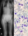
28 year old female patient presented to our clinic with normocytic anemia (hb: 6,50 g/dL), splenomegaly, short stature, limb and vertebral deformities. Clinically, the patient was evaluated for anemia. All routine blood investigations were done (Table 1). Bone marrow biopsy showed hypercellularity for age with increased rate in the erythroid series with marked dysmorphism findings. (figure 1) Next-generation sequencing was performed for diagnosis in the patient with dyserythropoiesis and morphological anomaly. Homozygous variant of CDIN1 gene was detected. Detailed NGS and karyotype analysis of the patient are shown in table 2 and 3. The patient diagnosed with congenital dyserythropoietic anemia type 1 and followed up with deferasirox and erythrocyte replacement.

Conclusion

CDAs are characterized by clinical and genetic heterogeneities. NGS based testing allows diagnosis. The increased knowledge of the genetic features and the detailed phenotyping of these patients will allow for the earliest start of the necessary treatment for the affected patients, as well as the monitoring of hemoglobin and iron levels. References:(1) Iolascon A, Andolfo I, Russo R. Congenital dyserythropoietic anemias. Blood. 2020 Sep 10;136(11):1274-1283. doi: 10.1182/blood.2019000948. PMID: 32702750. (2) Roy NBA, Babbs C. The pathogenesis, diagnosis and management of CDA type I. Br J Haematol. 2019. doi: 10.1111/bjh.15817. (3) Heimpel H, Kellermann K, Neuschwander N, Högel J, Schwarz K. The morphological diagnosis of congenital dyserythropoietic anemia: results of a quantitative analysis of peripheral blood and bone marrow cells. Haematologica. 2010;95(6):1034-1036

Texto Completo

Table 1.

Laboratory investigations

Laboratory Tests  Results  Normal Value 
Hemoglobin  6,50 g/dl  12,1 - 16,6 
Total Leukocyte Count  4,09 × 103/µl  3,46 - 10,04 
Platelets  232 × 103/µl  172 - 380 
Mean Corpuscular Volume  96,20 fl  81,8 - 98 
Reticulocytes  0,0747 × 106/µl  0,0188 - 0,1086 × 106/µl 
Total Bilirubin  1,15 mg/dl  < 1,2 
LDH  126  135-214 
İndrect Coombs  Negative 
Haptoglobulin  <0,1 g/l  0,3 - 2 

Figure 1: A) The patient's height was measured as 140 cm and scoliosis was detected in the skeletal survey. Bone marrow aspiration findings: B) Nuclear chromatin bridges between erythroblasts. C) Binucleate erythroblasts D) Multinuclear eryhtroblasts.

Table 2.

Results of NGS analysis: Homozygous variant of CDIN1 gene was detected. (AR: Autosomal recessive, CDIN1: congenital dyserythropoietic anemia type 1, PRF1: perforin 1, TAF6: TATA-Box Binding Protein Associated Factor 6)

Gene transcript  Position  Inheritence  Genotype 
CDIN1  Chromosome 15  AR  Homozygous 
PRF1  Chromosome 10  AR  Heterosygotic 
TAF6  Chromosome 7  AR  Heterosygotic 

Table 3.

Cytogenetic analysis of the patient: With the cytogenetic analysis of the patient, tetraploid chromosome formation was detected in 17 of 18 of the metaphases as 92XXXX. (DEB: clastogenic effect of diepoxybutane, G6PD: Glucose-6-Phosphate Dehydrogenase)

Chromosomal analysis  92,XXXX  TP53 deletion  %85 
Del20q12  %85  Del7q31.2  %83 
Del5q31.2  %85  Tetrasomy7/tetrasomy8  %85 
DEB  %0  G6PD  20,3 ıu/g Hb (normal) 
Pyruvate kinase  397,9 (normal)  Osmotic Fragility of Erythrocytes  Normal 

Baixar PDF
Idiomas
Hematology, Transfusion and Cell Therapy
Opções de artigo
Ferramentas