Acute myeloid leukemia (AML) is a neoplasm characterized by clonal proliferation and infiltration of bone marrow and other organs by poorly differentiated myeloid lineage cells. If left untreated, the outcome is usually fatal. It is the disease of the elderly in the western world; however, the median age at diagnosis in India is 40 years.1 AML accounts for 1.2 % of all cancer cases, highlighting that it is not a common malignancy. The patients with AML most commonly present with symptoms of pancytopenia, mainly easy fatigability, fatigue, bleeding manifestations or skin bruising, and fever. The uncommon presentations include Sweet syndrome, visual loss, gingival hypertrophy, palpable lymph nodes, hepatosplenomegaly, migratory arthritis, and myeloid sarcoma. Sudden visual loss due to central retinal vein occlusion (CRVO) is rarely reported as the initial presentation of AML, with only a few cases reported in the literature.2
Retinal venous occlusions can be classified on the basis of the site of occlusion as branch retinal vein occlusion (BRVO) which typically occurs at the cross-section of retinal artery and vein and central retinal vein occlusion (CRVO) typically occurs near the lamina cribrosa of the optic nerve. Further CRVO can be classified as ischemic and non-ischemic based on the degree of retinal capillary nonperfusion demonstrated after fluorescein angiography. Ischemic CRVO presents with poor visual acuity at initial presentation and poor visual prognosis in the long-term due to neovascularization.3 CRVO is a leading cause of sudden painless loss of vision and subsequent visual morbidity. In the age group of 40 - 49 years, the prevalence of CRVO is 0.06 %. The most common risk factors associated with CRVO are advanced age, hypertension, diabetes mellitus, stroke, coronary artery disease, female sex, raised creatinine and prothrombotic states.4 The management options for CRVO include anti-vascular endothelial growth factor(VEGF) therapy, glucocorticoid implants, and Laser therapy, with anti-VEGF therapy being the current first-line option.5
We present a case of a 40-year-old male with no relevant past medical history, who presented with sudden onset painless loss of vision in the right eye and, on evaluation, was found to have CRVO, pancytopenia, and azotemia. He was diagnosed as AML (intermediate-risk) and chronic kidney disease as a co-morbidity, and managed with hypomethylating agents.
Case reportA 47-year-old male with no significant past medical history, presented with complaints of painless diminution of vision in the right eye, first noticed on waking up from sleep. He did not have any burning, redness, or discharge from the right eye. He did not seek medical consultation for the next ten days. Subsequently, when his symptoms did not resolve, he consulted an ophthalmologist. On evaluation, he was found to have a distant visual acuity of 6/36 in the right eye and 6/9 in the left eye. His intraocular pressure was 14 and 13 mm of Hg in the right and left eye, respectively. Pupils were equal and reactive without an afferent pupillary defect, and extraocular movement, and confrontational visual fields were normal. The slit-lamp examination revealed unremarkable anterior chamber. On examination of the fundus of the right eye, there were blurred margins of the optic disc and multiple flame-shaped hemorrhages in all four quadrants, while the left eye was normal. Optical coherence tomography of the right eye showed central macular thickness of 758 micrometer and neurosensory retinal detachment. He was diagnosed as a case of central retinal vein occlusion right eye and was planned for intraocular anti–VEGF therapy. His initial laboratory evaluation revealed pancytopenia with a hemoglobin level of 5.2 g/dL, total leucocyte count of 2300/microL, and platelet count of 20000/microL. His mean corpuscular volume was 102 fL and a reticulocyte count of 1.92%. His initial serum creatinine was 5.4 mg/dL. However, urine routine examination and microscopic examination did not show any abnormality except proteinuria. His serum albumin level was 3.0 - 3.5g/dL on different occasions. The patient was reassessed, and the history was revisited. He denied any history of fever, ecchymotic patches, easy fatigability, increased or decreased urine output, blood in the urine, or frothy urine. On examination he had bilateral pitting pedal edema extending upto the knees; however he did not have anasarca. He was evaluated for the possible causes of pancytopenia. His initial peripheral blood smear showed macrocytic anemia with leucopenia and thrombocytopenia. Initially, the clinical diagnosis of Vitamin B-12 deficiency related pancytopenia was considered. He was started on injection methylcobalamin 1000 mcg intravenous once a day and oral folic acid supplementation empirically. However, his evaluation subsequently revealed normal serum vitamin-B12 and folate levels along with normal serum bilirubin, aspartate aminotransferase, alanine aminotransferase, and lactate dehydrogenase levels. The patient underwent bone marrow aspiration and biopsy, which showed diluted bone marrow aspirate. The bone marrow biopsy (BMBx) showed 60% cellularity with trilineage hematopoiesis with erythroid preponderance. Clustering of immature precursors away from the paratrabecular area was also noted. The bone marrow studies ruled out vitamin B-12 deficiency, and now the clinical diagnosis of pancytopenia with hypercellular marrow was made; however, acute leukemia, bone marrow infiltration, myelodysplastic syndrome, bone marrow failure were also ruled out. Simultaneously he was evaluated for the procoagulant states leading to CRVO. However, he was not found to have factor V Leiden and Prothrombin G20210A mutations. Protein C and Protein S deficiency were not detected. He was negative for antinuclear antibody, antiphospholipid antibody, anticardiolipin antibody, and antibodies against beta-2 glycoprotein. He also underwent ultrasonography of the abdomen, which showed a right kidney of 8.5 cm and left kidney of 9 cm, with bilateral raised echogenicity, and loss of corticomedullary differentiation. His 24 urinary protein was 3 grams. He underwent a repeat bone marrow aspiration and biopsy, that showed 30% blasts, which were myeloperoxidase positive (Figure 1a). Bone marrow biopsy showed clusters of blasts that were CD34 positive (Figure 1b, c). The flow cytometry confirmed the myeloid lineage as the blasts were CD13, CD33, MPO, CD34, CD117, CD45, and HLA-DR positive. He was diagnosed as a case of AML with intermediate-risk on the basis of cytogenetic abnormalities (Table 1) and was managed with Injection Decitabine 20 mg/m2 intravenous daily for five days every four weeks.

(A) Bone marrow aspitrate smear showing blasts with high N:C ratio (red arrows) along with an erythroid lineage cell showing dyspoietic changes (blue arrow) (MGG, 1000x). (B) Bone marrow biopsy showing hypocellalar marrow with clusters of blasts (H&E, 400x). (C) Immunohistochemistry for CD34 showing positivity in blasts (IHC, 400x).
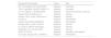
Cytogenetic abnormalities.
In acute myeloid leukemia, ocular signs are more common than visual symptoms. The most common visual symptom is the diminution of visual acuity. Rarely can a patient present with eye/ eyelid swelling due to infiltration of eye and surrounding structures by the leukemic cells.6 The ocular involvement is seen in 41-66 % of the cases. The most common ocular findings in AML are tortuous and dilated retinal veins, retinal hemorrhages, cotton wool spots, vitreous hemorrhages, papilledema, subconjunctival hemorrhages, eyelid swelling, optic disc edema, conjunctival infiltration, macular hemorrhages, subhyaloid hemorrhages, and rarely retinal vein occlusion.7,8 Out of these changes, the most common are intraretinal hemorrhages, white-centered hemorrhages, and dilated tortuous veins. The CRVO is very rare and was reported in only three patients out of 288 leukemia cases over seven years in a study by Reddy S C et al.7 Our patient presented with sudden painless loss of vision in one eye, and subsequently, he was found to have retinal hemorrhages in all four quadrants of the right eye and separation of the neurosensory retina from the retinal pigment epithelium.
Various hypotheses have been postulated regarding the exact cause of CRVO and BRVO in the patients with leukemia. The most common possible cause is hyperviscosity due to leukocytosis or thrombocytosis leading to leukostasis, vascular occlusion and hypoxic tissue damage. Other possible causes are concomitant infection, thrombosis or direct infiltration by the neoplastic cells. Recently Porte D L., described a case of 56-year-old who presented with CRAO in left eye and was diagnosed with leukemic relapse on optic nerve biopsy, even when there was no evidence of relapse on bone marrow studies, imaging, and CSF studies.9 Similarly microscopic infiltration by the malignant cells as the cause of bilateral retinal artery occlusion was described by Khair D et al., even when the patient had normal looking optic disc.10
Though our patient did not have hyperleukocytosis, it could have been the most likely explanation for CRVO of the right eye in our patient. However, retinal venous thrombosis or infiltration by malignant immature myeloid blasts (non-monocytic) with a tendency to adhere to the micro-vasculature due to the sluggish flow of the retinal veins may be the most likely explanation for CRVO in our patient. Acute monocytic leukemia can be associated with hyperviscosity syndrome, and renal insufficiency; however in our patient immunophenotyping did not show expression of monocytic differentiation antigens like CD14, CD4, CD11b, CD11c, CD64, CD68, CD36, lysozyme, and CD163.
AML could be 'de novo AML' or ‘secondary to MDS’ or ‘therapy-related AML’. Our patient was diagnosed with de novo AML as he had 30 % blasts on bone marrow aspirate smears and did not have a prior history of exposure to chemotherapeutic drugs, and he did not have cytogenetic abnormalities of MDS.
Immediately after the diagnosis of AML, the next step is risk stratification of the patient, which will further guide the therapy. The risk factors associated with a better or favorable outcome in AML are young age, good performance status (Karnofsky score > 60 percent), MDR-1 negative phenotype, no pre-existing hematologic disorders or prior chemotherapy, and favorable genetic mutations and karyotype. There is no clearly acceptable definition of younger in context of AML; however most of the studies take age > 50 years as older adults. The estimated five year overall survival rate varies with age ranging from 53 % in patients of 15 to 24 years to 3 % in patients 70 to 79 years old.11 This clearly highlights that the younger patients have better prognosis as was the case with our patient.
The patient is classified as an intermediate risk if there are no cytogenetic abnormalities classified as favorable or adverse [Table 1]. Our patient did not have any favorable or adverse cytogenetic abnormality; hence he was classified as intermediate risk.12
The current recommendation for the choice of induction therapy depends upon the risk category based on cytogenetic abnormalities and the risk of treatment related mortality (TRM). TRM is considered high, if the age is more than 70 years plus performance status 2-4 and /or any systemic co morbid condition like chronic kidney disease, chronic liver disease, heart failure, solid tumor with or without metastasis.12 Our patient was medically unfit but not frail and had a high risk of TRM as he had concomitant chronic kidney disease with the serum creatinine of 6.4 mg/dL and proteinuria of 3 gm/24 hours. Combination therapy including hypomethylating agent (HMA) along with Venetoclax has currently become the standard of care for medically unfit but not frail patients.13,14 However the initial treatment depends upon the availability of Venetoclax. In our case Venetoclax was not available at our centre as it is not yet licensed for use in India. So we managed our patient with HMA monotherapy: Injection Decitabine 20 mg/m2 intravenous per day for five days a month.
The chronic kidney disease was most likely due to presumed chronic interstitial nephritis as we could not biopsy the kidney in view of severe thrombocytopenia, and was likely discovered incidentally as an asymptomatic co-morbid illness in our patient. The CRVO of the right eye started improving; however renal function did not show any improvement after two cycles of hypomethylating agent (Decitabine) therapy.
ConclusionAML and CRVO are both uncommon disorders. The sudden loss of vision in one eye as the initial manifestation of AML is a rare occurrence. Our patient ignored it for ten days and did not seek medical attention. Incidentally, he was also found to have advanced azotemia and likely chronic interstitial nephritis leading to chronic kidney disease. Hypomethylating agents along with Venetoclax are the choice of therapy in medically unfit/ high risk for treatment related mortality patients.