

COVID-19 convalescent plasma is one of the experimental therapies used widely in moderately sick COVID-19 patients. However, there are a few risks involved in plasma transfusion; notably, transfusion-related acute lung injury (TRALI) caused by antibodies against human leukocyte antigens (HLA). This study was designed to assess the prevalence of anti-HLA antibodies in convalescent plasma donors using the single antigen bead method.
Study design and methodsThis was a hospital-based observational study of consecutive plasma donors. A total of 252 samples were screened for anti-HLA Class I and Class II antibodies using the microbead assay with the identification of anti-HLA Ab in positive samples being performed using a single antigen bead assay. Luminex-based normalized background cutoff ratios of 10.8 for Class I and 6.9 for Class II and mean fluorescence intensity cutoffs of 2500 for Class I and 1500 for Class II were used for screening and the single bead assay, respectively.
ResultsOf 252 screened samples, 28 (11.1 %) were positive for Class I, Class II or both Class I and Class II anti-HLA antibodies in donors with no history of a previous immunizing event. Moreover, 20/252 (7.9%) donors without any history of prior immunization had specific anti-HLA antibodies of Class I or Class II or both by the single bead assay.
ConclusionsThe high prevalence of anti-HLA antibodies in our cohort of donors raises an urgent and immediate need for anti-HLA antibody screening in all convalescent plasma donors for safe therapy of COVID-19 patients.
Emerging outbreaks of acute respiratory syndrome coronavirus-2 (SARS-CoV-2) infection have become a challenging issue of national and international concern. Coronavirus Disease (COVID-19) is an emerging infectious disease with a dramatic rise in global incidence, including in India.1 Symptoms of COVID-19 disease are non-specific; the disease presentation can range from no symptoms to severe pneumonia and death.2 The new SARS-CoV-2 epidemic is classically associated with respiratory illness and a few extra-pulmonary signs in patients with moderate and severe disease. COVID-19 is caused by severe SARS-CoV-2 and has been the most discussed disease in recent times (www.who.int). Antibody responses from diverse SARS-CoV-2 infections are vividly reported, even as the comorbidities or associated phenotypes emerging from the disease are homogenous, allowing us to ascertain relationships between homogeneity and heterogeneity of susceptible patients. In particular, the human leukocyte antigen (HLA) antibodies have been of inherent use in the emergence of SARS-CoV-2 infection. Their reaction with white blood cells further infects the lungs as plasma carries the highest risk of transfusion-related acute lung injury (TRALI) infection, indicating that HLAs match the infection prototype. Over the years, TRALI-related deaths has been reported worldwide.
COVID-19 Convalescent Plasma (CCP) is one of the experimental therapies being widely used in COVID-19 sick patients across the globe.3 There are reports that CCP may reduce the severity of the disease, but the therapy also has certain risks. For example, one of the most serious risk factors of plasma transfusion is antibody-mediated TRALI.4,5 Antibodies cause TRALI against human HLAs.6 However, the risk is of more significant concern since symptoms of TRALI may be masked by the natural course of the COVID-19disease, where respiratory symptoms are common. Interestingly, TRALI and COVID-19 share similarities in the pro-inflammatory respiratory scenario, e.g., neutrophil priming, elevated cytokine levels, and the formation of neutrophil extracellular traps (NETs) indicate that neutrophils might play a key role in both TRALI and COVID-19. 7 Critically ill COVID-19 patients may have a particular risk for developing single-donor plasma-induced TRALI and so TRALI prevention by anti-HLA antibody testing of convalescent single-donor plasma appears essential.
Plasma therapy has been used as an early emergency intervention in many outbreaks, including in Spanish Flu, severe acute respiratory syndrome (SARS), Middle East respiratory syndrome (MERS), and Ebola.8 In the present epidemic of COVID-19, COVID CCP is being collected by apheresis from people who have recovered from COVID-19. Plasmapheresis is a medical procedure where whole blood is removed from a donor (Recovered COVID-19 patient) and separated into individual components. Only one component (plasma) can be harvested for therapeutic purposes. As the CCP contains protective, neutralising antibodies of coronavirus, these antibodies have a direct antiviral action by suppressing virus replication in sick patients, thereby reducing mortality. The presence of anti-inflammatory cytokines, clotting factors, natural antibodies, and other undefined proteins in the plasma may further enhance anti-inflammatory and immune modulator response.9 CCP can be collected 14 days after complete COVID-19 recovery. Anyone between 18 and 60 years who has recovered from COVID-19 is eligible to donate their blood plasma, even without a negative reverse transcription polymerase chain reaction (RT-PCR) report 14 days after the cessation of symptoms, completion of treatment, or home isolation.10
TRALI is a severe pulmonary syndrome that can sometimes lead to death if not recognised and treated. Patients who receive plasma-containing products may be at risk for TRALI; it is consistently a leading cause of transfusion-related mortality; it is fatal in 5–10 % of all cases.11,12 COVID-19 patients with acute respiratory distress syndrome (ARDS) or who could progress to ARDS may have a higher risk of TRALI due to their underlying lung injury. Clinical and preclinical studies have shown that an inflammatory ‘first hit’ is almost always present before TRALI onset. Some clinical studies have identified ARDS risk factors that may also be TRALI.13,14 To ensure a COVID patient receiving CCP does not develop TRALI, the plasma should be screened for anti-HLA Class I and anti-HLA Class II antibodies. Class I anti-HLA specific antibodies can bind donor white blood cells directly to the lung endothelium, leading to neutrophil activation. Class II anti-HLA specific antibodies are thought to bind to major histocompatibility complex Class II expressed on recipient monocytes, thereby activating neutrophils.
Therefore, there is an urgent need to check for the prevalence of anti-HLA antibodies in Recovered COVID-19 plasma donors. Prevention strategies like testing for anti-HLA antibodies in CCP donors have not been studied in India as yet. Therefore, the present study was planned to assess the prevalence and specificities of anti-HLA antibodies in CCP donors using microbead-based single antigen bead (SAB) assays on the Luminex platform.
Material and methodsStudy design and populationThis was a hospital-based observational study from two different plasma collection blood centers of north India from August 1 to November 30, 2020, after approval from the Institutional Ethics Committee, Mahatma Gandhi University of Medical Sciences and Technology, Jaipur. CCP donors were eligible for inclusion irrespective of gender (parous females were excluded) as per national guidelines and criteria laid down by the Indian Society of Transfusion Medicine (ISTM) in the guidance document for CCP donation.10 Only samples of contemporary CCP first-time donors, who gave no history of previous immunization events like a transfusion, were included in the study. If the donor came back for repeat apheresis donations, their sample was excluded from the study.
After giving informed consent, CCP donors completed the CCP apheresis questionnaire form regarding the status of the RT-PCR test for COVID-19 disease, number of days from the resolution of last symptoms, and prior immunizing events. In addition, an 8-mL serum sample was obtained for the anti-HLA antibody analysis additional to the routine complete blood count, transmissible transfusion infections testing, serum protein estimation, and anti-SARS CoV2 IgG antibody testing.
Sample preparation and testingSerum samples collected from CCP donors were centrifuged for ten minutes at 5000 g and stored at −40 °C until further analysis. Batch testing for anti-HLA Class I and Class II antibodies were performed with the microbead-based assay technique (Luminex, Austin, TX, USA) using LAB Screen™ Mixed Class I & II (One Lambda, Canoga Park, CA) to screen anti-HLA Class I and Class II antibodies. Positive samples by the LABScreen™ Mixed screening test using normalized background (NBG) ratios >10.8 for Class I and >6.9 for Class II were further tested with LABScreen™ Single Antigen HLA Class I and Class II (One Lambda, Canoga Park, CA) testing for anti-HLA antibody specificity.
Study endpointThe study's primary outcomes were the prevalence and specificities of anti-leukocyte antibodies in CCP donors, who did not have any history of previous immunizing events like pregnancy, transfusion, or transplant. LabScreen™ Mixed Class I and II screened samples and LABScreen™ single antigen HLA Class I and Class II specificities were analyzed for all positive filtered samples.
ResultsA total of 252 samples were screened for anti-HLA Class I and Class II antibodies using the Labscreen mixed Class I/II test. Specific anti-HLA Ab positive filtered samples were confirmed by a SAB assay using Luminex-based NBG cutoff ratio and mean fluorescence intensity (MFI), respectively. The donor characteristics and results of anti-HLA antibody testing are reported. In the present observational study, 252 CCP donors who never had any potentially immunizing events were screened for anti-HLA antibodies. Of the 252 donors, 240 were male and 12 were female. The mean age of the CCP donors was 34.5 years and the average number of days from the cessation of symptoms to sample collection was 43.9 days.
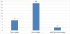
Of the 252 samples, the microbead mixed antigen assay detected 28 (11.11 %) donors with the anti-HLA Class I and Class II antibodies. (Figure 1)

Of the 28 positive samples, seven (25 %) were positive for Class I, 19 (67.8 %) were positive for Class II, and two (7.14 %) samples were positive for both considering NBG cutoff values of 10.8 and 6.9 for Class I and Class II, respectively based on the previous study by Endres et al. 16 All 28 positive donors were male donors (Figure 2).

For data analysis, the two samples that tested positive for both HLA Class I and Class II antibodies were included as both in Class I and Class II positive samples.
Using the Class I SAB cutoff of 2500 MFI, 77.7% (7/9) of the serum samples positive in the screening assay were positive by the Class I SAB assay, and 63.1 % (12/19) that tested negative in the screening assay were negative in the SAB assay. Labscreen Mixed test to identify HLA Class I positive samples had sensitivity of 50 %, specificity 85.71 %, positive predictive value 77.77 % and negative predictive value 63.15 %. The sensitivity and specificity of the Labscreen mixed test is based on the leukocyte Antibody Prevalence study (LAPS) carried out by Endres et al. (16) using a Luminex-based NBG cutoff ratio of 10.8 (Class I). (Table 1)
Using the Class II SAB cutoff of 1500 MFI, 52.3 % (11/21) of the serum samples that tested positive in the screening assay tested positive in the SAB assay, and 28.7 % (2/7) of samples tested negative in the screening assay were also negative in the SAB assay. Labscreen Mixed test to identify HLA Class II positive samples had sensitivity of 68.75 %, specificity 16.66 %, positive predictive value 52.38 % and negative predictive value 28.57 %. The sensitivity and specificity of Labscreen mixed test is based on the leukocyte antibody prevalence study (LAPS) performed by Endres et al. (16) using the Luminex-based NBG cut-off ratio of 6.9 (Class II - Table 2).
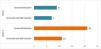
Of the nine samples positive for anti-HLA Class I antibodies in the Labscreen Mixed test, seven were positive by the SAB assay. Of the 21 samples positive for anti-HLA Class II antibodies in the Labscreen Mixed test, 11 were positive by the SAB assay (Figure 3).
Discussion
This study demonstrated that 7.9% of CCP donors have Class I or Class II anti-HLA antibodies or both by SAB assay. In the present observational study, 252 CCP donors who never had any potentially immunizing events were screened for anti-HLA antibodies. The microbead mixed antigen assay detected 28 (11.1 %) donors with Class I, II, or anti-HLA antibodies. Out of 28 positive samples, seven (25 %) were positive for Class I, 19 (67.8 %) were positive for Class II, and two (7.14 %) samples positive for both considering NBG cutoff values of 10.8 for Class I, and 6.9 for Class II, and MFI >2500 (Class I) and >1500 (Class II) as cutoff values for the specificity of anti-HLA by SAB assay based on the LAPS study by Endres et al. 17 In the current cohort, 95% of CCP donors were male, and 5 % were female. Kleinman et al. found anti-HLA Class I and Class II antibodies to cognate antigens in TRALI cases applying MFI cutoffs >2500 for type I and >1500 for Class II.11 In a prospective surveillance study, cognate anti-HLA Class II antibodies with MFI >1500 were associated with TRALI. In contrast, cognate anti-HLA Class II antibodies with MFI of 300–1500 did not pose a substantial TRALI risk. Plasma from donors with anti-HLA antibodies should not be prepared to prevent TRALI.15 Concerning CCP, the provision of male or nulliparous female donor CCP has been adopted by various national transfusion services, including India. However, screening for anti-HLA antibodies and excluding antibody-positive donors is a more focused approach suitable for CCP donations.
Other studies on plateletpheresis donors have demonstrated the feasibility of SAB assays for anti-HLA antibody screening. 17,18 Data suggest that the presence of a specific anti-HLA antibody in plasma products causes the development of TRALI correlated with the strength of MFI.12,19 However, using SAB assays for anti-HLA antibody testing has various limitations: variable testing procedures and non-standardized test kits lead to inter-laboratory variations. 20 In addition, no consensus cutoff for donor screening for TRALI prevention exists. NBG cutoffs range from 22 to 59.3 for anti-HLA Class I and 27.5 for anti-HLA Class II antibodies for mixed antigen assays. 17,21 Similarly, single antigen cutoffs vary from MFI >1000 for both anti-HLA type I and Class II antibodies to MFI >2500 for type I and MFI >1500 for Class II antibodies.11,16,22 These varying cutoffs reflect the different approaches to cut off determination. Some authors chose cutoffs as suggested by the manufacturer. Others based them on internal laboratory validation data or tailored cutoffs for the specific donor population tested considering non-transfused males and never-pregnant females.22,23
In the recent past, various studies confirmed the presence of anti-HLA antibodies in never exposed donors. In the current study, 7.9% of male donors were positive compared to 4.3 %, which is a significant finding.16,24,25 However, the clinical relevance of these antibodies has been a matter of debate. 26 A study of CCP donors by Juskewitch et al. shows the presence of specific anti-HLA antibodies in 7.2% of males never exposed to previous immunizing events, which correlates with the present findings. 27 The current COVID-19 pandemic emphasizes the relevance of TRALI prevention by anti-HLA antibody testing, as CCP transfusions in the treatment of COVID-19 may pose an additional risk, mainly when CCP containing anti-HLA antibodies is transfused.
On the other hand, TRALI is thought to be a two-step process. Under normal conditions, the lung vasculature still has substantial numbers of neutrophils. These neutrophils can become partially activated by pulmonary tissue inflammation (usually of unknown origin but in COVID patients, this may occur due to the hallmark-associated cytokine storm) but may remain below the threshold for TRALI development. A second trigger is required for TRALI to develop; this triggers the transfer of antibodies from various transfusion products to the patient. Due to high plasma volume, fresh frozen plasma, platelets, and packed red blood cells have all been implicated with TRALI. In the COVID setting, this second trigger may occur due to the administration of CCP.
Some discrepancies between the screening and SAB assays were observed in the present study. For example, 22.2 % of samples with positive Class I screening results had negative SAB results, and 47.6 % of samples with positive Class II screening results had negative SAB results. This reactivity could be due to multiple low-titer antibody specificities binding to the same individual screening beads that were coated with antigens from various cell lines or due to false-positive screening test results. We also observed the opposite type of discrepancy, 36.8 % of samples that screened negative by the cutoff of NBG (10.8) for Class I were positive by SAB assay, and 71.4% of the sample that screened negative by cut off of NBG (6.8) for Class II were positive by the SAB assay. The higher percentage of positivity on SAB is possibly due to higher antigen density on the SAB beads and higher cutoff based on the LAPS study in contrast to manufacturers recommendation of NBG cutoff value of >2.2 for Class I and Class II.
As a part of national guidelines for CCP, blood centers are currently only using plasma from male donors and nulliparous female donors. However, as a patient of COVID-19 already has acute lung injury, giving anti-HLA antibody-positive CCP may lead to a fatal TRALI reaction, which the natural progression of the disease can mask. This study aimed to check the prevalence of anti-HLA antibodies in CCP donors. We argue that our research has demonstrated that 7.9% of CCP donors have the specific anti-HLA antibody. However, they did not have any prior immunisation history except COVID-19 the disease compared to 4.3 % anti-HLA antibody in regular healthy blood donors by sensitive bead-based assays methods.27
ConclusionsSARS-CoV-2 infections have been on the rise, and there is an immediate need for understanding the exact clinical significance of the presence of anti-HLA antibodies. In this study, we highlighted the scope and potentiality of CCP donors having a higher positivity rate when compared to regular healthy donors. Moreover, positivity in males suggests that all the blood center facilities involved in CCP collection should screen for anti-HLA antibodies in eligible CCP donors. Therefore, deferring such donations would mitigate the risk of TRALI in COVID-19 disease recipients. Our study would also recommend standard operating procedures (SOPs) for future anti-HLA donor studies.
CRediT authorship contribution statementRam Mohan Jaiswal: Conceptualization, Data curation, Formal analysis, Funding acquisition, Investigation, Methodology, Project administration, Resources, Writing – original draft, Writing – review & editing. Aseem Kumar Tiwari: Formal analysis, Writing – original draft, Writing – review & editing. Ashina Singla: Software, Supervision, Validation, Visualization. Ashish Jain: Software, Supervision, Validation, Visualization. Rajaat Vohra: Formal analysis, Funding acquisition, Investigation, Methodology, Project administration, Resources, Software, Supervision, Validation, Visualization. Munish Kakkar: Formal analysis, Writing – original draft, Writing – review & editing. Prashanth Suravajhala: Software, Supervision, Validation, Visualization, Writing – original draft, Writing – review & editing.