


Consensus of the Brazilian association of hematology, hemotherapy and cellular therapy on patient blood management
Mais dadosManaging coagulation disorders and potential bleeding risks, especially in the context of anticoagulant medications, is of immense value both clinically and prior to surgery. Coagulation disorders can lead to bleeding complications, affecting patient safety and surgical outcomes. The use of Patient Blood Management protocols offers a comprehensive, evidence-based approach that effectively addresses these challenges. The problem is to find a delicate balance between preventing thromboembolic events (blood clots) and reducing the risk of bleeding. Anticoagulant medications, although crucial to preventing clot formation, can increase the potential for bleeding during surgical procedures. Patient blood management protocols aim to optimize patient outcomes by minimizing blood loss and unnecessary transfusions.
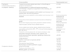
Thrombocytopenia, defined as a platelet count below 150 × 109/L, occurs in approximately 5–10% of patients in the preoperative period. An individual's baseline platelet count is relatively stable throughout life, so an important change in this count may be due to a physiological change, as occurs during pregnancy, or to a pathological process. Although hemostatic function depends on multiple factors, platelet number and function are key elements.1 A platelet transfusion should not be indicated in elective surgery patients with thrombocytopenia for whom the etiology is unclear, before elucidating the cause.2Table 1 summarizes the main mechanisms related to thrombocytopenia that must be considered in the preoperative period.
Main differential diagnosis of thrombocytopenia.
Adapted from Estcourt et al. (2017).2
Medications such as antiplatelets, non-steroidal anti-inflammatory drugs (NSAIDs), anticonvulsants (antiepileptics) and antidepressants can negatively affect platelet function. Uremia, sepsis (platelet dysfunction induced by bacteria) and, of course, hereditary platelet diseases can also impair its function. Therefore, the preoperative clinical history of bleeding is essential and will identify whether it is necessary to continue the investigation before surgery.
For surgical purposes, thrombocytopenia can be characterized as mild (100–149 × 109/L), moderate (50–100 × 109/L) and severe (<50 × 109/L). Despite this classification, it is known that the relationship between the platelet count and the risk of bleeding is not linear and that the hemostatic capacity depends on other variables. Studies suggest that the risk of spontaneous bleeding is difficult to predict until the platelet count reaches very low values (<10 × 109/L). In addition to platelet count, many other factors affect the likelihood of bleeding, including the cause of thrombocytopenia, the patient's age, as well as the presence of concomitant infection/sepsis and medication use. All these variables must be considered before recommending a platelet transfusion.3
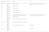
There are few data to guide the moment to prescribe a platelet transfusion in surgical procedures. Recommendations are mostly based on studies with weak evidence and on expert opinions. The greatest experience comes from studies involving oncology and hematology patients. Platelet transfusions may be indicated for bleeding prophylaxis in patients with very low counts (and presence of risk factors for major bleeding) and before invasive procedures, or as therapy for the treatment of active bleeding. The prompts for indicating prophylactic platelet transfusion vary according to the procedure and, often, there is no consensus among experts. Table 2 summarizes some of the indications for prophylactic transfusion.4
Recommended platelet count in different clinical conditions.
| Clinical condition | Recommended count | |
|---|---|---|
| Prophylactic indication(without bleeding or justgrade 1 bleeding) | Transient bone marrow aplasia secondary to chemotherapy or allogeneic transplantation | ≥10 000 |
| Thrombocytopenia in a patient with aplasia secondary to chemotherapy or allogeneic transplantation without bleeding, but with risk factora | ≥20 000 | |
| Chronic bone marrow aplasia in which platelet count recovery is not expected after intensive drug treatment | ≥10 000 | |
| Chronic bone marrow aplasia in which platelet count recovery is not expected | Routine prophylactic transfusion not recommended | |
| Chronic bone marrow aplasia to prevent recurrence of bleeding ≥ grade 2 | Individually evaluate each case | |
| Bone marrow biopsy and puncture, peripheral catheter insertion, cataract surgery, central venous catheter removal | Routine prophylactic transfusion not recommended | |
| Chronic stable bone marrow aplasia, platelet function disorder, consumption or destruction (DIC, PTT), immune thrombocytopenia (PTI, HIT, PPT) | prophylactic transfusion is not indicated | |
| Transient or chronic aplasia with risk factors (critically ill, other associated coagulation disorders, sepsis, antibiotic use) | 10 000 – 20 000 | |
| Central venous catheter implantation (guided by ultrasound by experienced team) | >20 000 | |
| Lumbar puncture | ≥ 40 000 | |
| Insertion or removal of an epidural catheter | ≥80 000 | |
| Major surgery | >50 000 | |
| Percutaneous hepatic biopsy | >50 000 | |
| Neurosurgery or posterior ophthalmic surgery | >100 000 | |
| Therapeutic indication | Severe hemorrhage | >50 000 |
| Polytrauma, brain lesion, CVA | >100 000 | |
| Bleeding grade ≥ 2, not severe | >30 000 |
DIC: Disseminated intravascular coagulation; TTP: Thrombotic thrombocytopenic purpura; HIT: Heparin-induced thrombocytopenia; ITP: Immune thrombocytopenia purpura; PPT: Periparturient thrombocytopenia; CVA: Hemorrhagic cerebrovascular accident.
consider not performing prophylaxis in autologous transplantations
Adapted from Estcourt et al. (2017).2
Other non-transfusion measures can be considered to control bleeding associated with thrombocytopenia. The use of antifibrinolytics in patients with thrombocytopenia due to bone marrow failure can reduce bleeding and the need for transfusions. They are also used to control bleeding in patients with platelet dysfunction and should be indicated in cases of hemorrhage associated with trauma and in patients who are candidates for major surgery that are expected to lose >500 mL. The use of DDAVP (desmopressin) may be useful in the treatment of bleeding in patients with suspected platelet dysfunction due to uremia as it increases the release of Von Willebrand multimers by the endothelium, but it is effective for a short period of time as it exhibits tachyphylaxis after the first doses. It can also be used in inherited platelet disorders to control minor bleeding, generally associated with an antifibrinolytic.2,5 More details about antifibrinolytics are considered in an article that is part of this consensus.
Studies with thrombopoietin (TPO) receptor agonists show that this class of medications can be an alternative to improve platelet counts before major surgical procedures for different pathologies such as hematological diseases, immune thrombocytopenia, chemotherapy toxicity and storage disease and especially in chronic liver disease. Despite reducing the need for platelet transfusions and the associated risks (allergic reactions, alloimmunization/platelet refractoriness, transfusion-related acute lung injury - TRALI, sepsis, etc.), the cost of the drug still limits its routine use.6,7
With the exception of the use of factor VIIa, there is no evidence that favors the use of bypass agents such as Factor VIIr, prothrombin complex concentrate (PCC) and activated PCC in the prophylaxis or treatment of bleeding in patients with thrombocytopenia. Factor VIIa can be indicated in more severe cases of congenital bleeding disorders (Glanzmann's Thrombasthenia).1,2
Management of platelet antiaggregation in elective proceduresThe decision to suspend antiplatelet therapy must take into account the potential risk of bleeding during the surgical procedure and, on the other hand, the risk of thrombotic events considering the reason that antiplatelet therapy was initially prescribed. The pharmacological properties of each antiplatelet drug will dictate the moment of interruption and its resumption in the postoperative period. Acetylsalicylic acid (AAS) and the P2Y12 inhibitors, clopidogrel and prasugrel, irreversibly inhibit platelet function and, when necessary, suspension must occur sooner to allow platelet function to be restored. The P2Y12 inhibitor ticagrelor, on the other hand, reversibly inhibits platelet function but requires interruption at least two to three days before the surgery.8
AAS: For patients taking AAS who will undergo an elective surgery, it is recommended that the antiplatelet agent be maintained, including in cardiac surgeries. Suspension should be considered when AAS is used as primary prophylaxis and in cases with a high risk of bleeding, such as neurosurgery, with suspension being suggested five days before the procedure.8,9 For minor procedures such as tooth extraction, dermatological and ophthalmological procedures, routine suspension of AAS is not recommended.8
P2Y12 inhibitors: The suspension of P2Y12 inhibitors is suggested for patients who will undergo cardiac and non-cardiac surgeries. It is recommended to suspend clopidogrel five days in advance, ticagrelor three to five days in advance and prasugrel seven days in advance.8 For minor procedures such as tooth extraction, dermatological and ophthalmological procedures, the routine suspension of P2Y12 inhibitors is not recommended.
Dual antiplatelet therapy: For patients with coronary artery disease with recent stent implantation who will undergo surgery, suspension of dual antiplatelet therapy (AAS + P2Y12 inhibitor) is not recommended. When possible, the surgical procedure should be postponed. If the stent was placed within the 12 previous weeks, it is recommended to maintain dual antiplatelet therapy or suspend just one antiplatelet agent. If the stent implantation took place over three months before, it is recommended to suspend the P2Y12 inhibitor. The decision must involve the cardiologist as several factors must be considered: the type of stent (whether drug-eluting or not), the time after stent placement, the number and location of implants. Routine suspension of double aggregation using bridging with a IIb/IIIa inhibitor (cangrelor) or a low molecular weight heparin (LMWH) is not recommended. Furthermore, for patients using double aggregation who will undergo procedures with a low risk of bleeding, it is suggested to suspend only the P2Y12 inhibitor.8
Platelet function tests: In patients receiving antiplatelet drug therapy who will undergo an elective procedure, the routine use of preoperative platelet function tests to guide preoperative antiplatelet management is not currently recommended. It may possibly be beneficial to use platelet function testing in certain scenarios, such as patients undergoing coronary artery bypass graft (CABG) surgery who have recently started taking a P2Y12 inhibitor, but the considerable costs of testing must be taken into account.8
The patient should also be asked about the use of other less commonly used platelet function inhibitors, which should also be discontinued. These drugs include cilostazol, dipyridamole and pentoxifylline with half-lives of 2 to 10 h. Nonsteroidal anti-inflammatory drugs have reversible antiplatelet properties with half-lives ranging from 2 to 6 h (ibuprofen, ketoprofen, indomethacin), from 7 to 15 h (celecoxib, naproxen) to approximately 20 h (meloxicam, piroxicam).
Figure 1 summarizes the preoperative approach to antiplatelet agents.
 *It is recommended to maintain the drug. Consider suspension in procedures with high risk of bleeding, such as neurosurgery. **Bridging with cangrelor is not routinely indicated, however, it should be considered in patients at high risk of thrombosis and should begin 72 h after discontinuation of the P2Y12 inhibitor. ***P2Y12 inhibitors must be started within 24 h after the procedure at the maintenance dose. †The suspension must occur 3 to 5 days before the procedure. ‡The suspension must occur 5 days before the procedure #suspension must occur 7 to 10 days before the procedure. Adapted from Douketis et al. (2022).8.")
Preoperative approach to antiplatelet drugs (antiaggregants)
*It is recommended to maintain the drug. Consider suspension in procedures with high risk of bleeding, such as neurosurgery. **Bridging with cangrelor is not routinely indicated, however, it should be considered in patients at high risk of thrombosis and should begin 72 h after discontinuation of the P2Y12 inhibitor. ***P2Y12 inhibitors must be started within 24 h after the procedure at the maintenance dose. †The suspension must occur 3 to 5 days before the procedure. ‡The suspension must occur 5 days before the procedure #suspension must occur 7 to 10 days before the procedure. Adapted from Douketis et al. (2022).8.
The preoperative management of oral anticoagulants (OAC) will depend on the characteristics of the OAC, the risk of bleeding associated with the procedure and the patient, as well as the risk factors for thromboembolic events. The risk of bleeding in each procedure should determine whether the OAC should be discontinued, when it should be discontinued and when it should be restarted postoperatively. In parallel, the risk of thromboembolic events will indicate whether it is necessary to change to unfractionated heparin or LMWH to minimize the risk of a new event in the preoperative period.10
Procedures must be classified as having minimal bleeding risk, low-moderate risk or high risk (Table 3) and the risk of venous thromboembolism must be estimated for the patient (Table 4). It is recommended that services use standardized tools to stratify the risk of bleeding and venous thromboembolism (VTE).
Bleeding risk stratification depending on the procedure.
Adapted from Spyropoulos et al. (2019).10
Stratification of preoperative thromboembolic risk.
a) Pancreatic cancer, active myeloproliferative disease, brain tumor, gastric cancer. b) Previous arterial fibrillation, stroke, transient ischemic attack, hypertension, diabetes, heart failure, age >75 years. c)History of oncological disease except non-melanoma skin lesions. VTE: Venous thromboembolism.
Adapted from Spyropoulos et al. (2019).10
The clinical approach will depend on the anticoagulant in use, whether vitamin K antagonist (VKA) or direct oral anticoagulants (DOAC). In general, stopping oral anticoagulants (OAs) is not routinely recommended for procedures with minimal risk of bleeding. For procedures with higher risk, OAs should be interrupted. In the case of DOACs, bridging with heparin is not recommended because the half-life of these drugs is short allowing suspension close to the procedure. For patients using warfarin, bridging can be considered in patients at high or intermediate risk of VTE, but it is not suggested in patients at low risk of VTE (Table 5).11
Oral anticoagulant management according to the bleeding risk and preoperative venous thromboembolism.
OAC: Oral anticoagulant; VKA: Vitamin K antagonist; DOAC: Direct oral anticoagulant; LMWH: Low molecular weight heparin; (a) In arterial fibrillation consider bridge for patients with high-risk CHADS2 (Scores 5 and 6). (b) Consider prophylactic dose of LMWH in patients submitted to procedures with high risk of bleeding or major surgeries associated with a high risk of venous thromboembolism. (c) Consider suspending the DOAC only on the day of the procedure.
Adapted from Spyropoulos et al. (2016).11
When bridging with heparin is indicated, the schedule should involve performing an international normalized ratio test (INR) seven days before the procedure, suspending the drug (D-6 to d-5), introducing LMWH on d-3 and repeating the INR on d-1 to assess the need for additional vitamin K (Table 6).
Preoperative management of warfarin with bridge of Low molecular weight heparin.
LMWH: Low molecular weight heparin; INR: international normalized ratio test; VKA: Vitamin K antagonist; VO: Via oral.
Adapted from Spyropoulos et al. (2016).11.
The timing of the discontinuation of DOACs, as well as their reintroduction, will depend on the risk of the procedure and the patient's renal function (Table 7).
Preoperative management of direct anticoagulants.
(a) Includes any surgery involving neuraxial anesthesia. (b) For patients with high risk of venous thromboembolism, consider reintroduce reduced dose of dabigatran (755 mg BID), rivaroxaban (10 mg/day) or apixaban (2.5 mg BID) in the afternoon after surgery and in the first postoperative. (c) Value considered for patients receiving 15 mg/day of rivaroxaban.
Adapted from Spyropoulos et al. (2016).11
Situations involving severe bleeding associated with OACs, hemorrhagic stroke or cases of urgent/emergency surgery may require rapid reversal of anticoagulation. It is essential that every service has established a protocol for reversal in accordance with the anticoagulant therapy in use.
Reversal of vitamin K antagonistsEven when administered intravenously, vitamin K can take hours to reverse OACs, as it depends on the production of factors II, VII, IX and X by the liver. This makes its use inappropriate as the solitary reversal strategy in the face of serious and life-threatening hemorrhages. Historically, fresh frozen plasma (FFP) was widely applied to reverse vitamin K antagonists, but its efficacy in the emergency setting has never been established. Furthermore, the use of plasma implies a delay in starting therapy due to the need for ABO typing and thawing. Another limiting factor is the volume of plasma required for reversal, which is generally high and may require a longer infusion time. As it is a blood component, the patient is exposed to the risk of transfusion reactions, such as volume overload, TRALI, transmission of infectious diseases, allergic reactions, etc.
PCC was initially developed to treat patients with hereditary deficiencies of coagulation factors, especially patients with hemophilia A and B. However, over time it began to be used to reverse the action of vitamin K antagonists and was approved by the FDA in 2013 for this purpose. In studies, PCC reversed the INR more quickly than FFP and its clinical response was not inferior. Furthermore, the occurrence of thromboembolic events was not higher with PCC. Therefore, currently PCC, associated with intravenous vitamin K, is considered the first choice for the urgent reversal of vitamin K antagonists.9
Reversal of direct oral anticoagulantsExperience with DOACs, prior to the development of their antidote, shows that many patients with bleeding associated with these drugs do not require reversal agents and can be controlled with supportive measures alone. Such measures include discontinuation of the DOAC and other medications known to interfere with hemostasis, local compression or direct treatment of the bleeding site, fluid replacement and, when necessary, transfusion support. Patients with mucosal bleeding may benefit from antifibrinolytic therapy. Oral activated charcoal can be used to remove unabsorbed DOAC from the gastrointestinal tract, particularly if it has been taken within the previous few hours. Therefore, reversal agents should be reserved for life-threatening bleeding, bleeding at a critical site such as the central nervous system, or major bleeding that is unresponsive to the above-mentioned supportive measures.12
When considering a reversal agent, it is important to assess the degree of anticoagulation and the likelihood that the anticoagulant is contributing to the bleeding. If assays to measure plasma DOAC concentration are not available, the degree of anticoagulation can be estimated based on the specific agent, dose, interval since last dose, and renal and hepatic function. Thus, the use of a reversal agent should only be considered when the estimated serum level of DOAC is clinically relevant.12
Two specific reversal agents have been approved by the Food and Drug Administration (FDA) in the USA: idarucizumab to reverse dabigatran and andexanet alfa (inactivated recombinant factor Xa) to reverse apixaban and rivaroxaban. The use of PCC is considered off-label and controversial for the treatment of severe bleeding in the absence of specific DOAC inhibitors.
Preoperative management of hereditary coagulopathiesPreoperatively, patients with hereditary coagulopathies should be evaluated together with the hematologist who monitors them. In suspected, but unconfirmed, cases of hereditary coagulation disorders, it is recommended that surgery be postponed until the diagnosis is completed, allowing for targeted management of each specific disorder. Von Willebrand disease (VWD) and hemophilia (factor VIII or IX deficiency) are the most common hereditary coagulopathies.13,14 Deficiencies of factors I (fibrinogen), II (prothrombin), V, VII, X and XIII are considered rare coagulopathies.15
It is important to highlight that the use of blood components (FFP or cryoprecipitate) should not be indicated for the correction of hereditary deficiencies of coagulation factors for which blood products containing specific factors are available (Factor VIII, IX, XI, XIII concentrates, Platelet components, apheresis platelet concentrate, Factor VIII and Von Willebrand factor, fibrinogen) or even recombinant factors (Factor VIIr, Factor VIIr).13-15
Indication of other possible situations that affect coagulationThe doctor must evaluate other conditions that affect a patient's coagulation and that are not directly linked to hereditary coagulopathies or to the use of DOACs, platelet aggregation inhibitors and antiplatelet agents, such as:
*Liver diseases: Liver disorders, such as cirrhosis and hepatitis, can interfere in the liver's production of clotting proteins, such as the synthesis of clotting factors. This can result in an imbalance in the clotting cascade and result in abnormal bleeding.
*Hepatorenal syndrome: In this condition, kidney function is affected due to severe liver diseases. This can lead to clotting disorders resulting from kidney failure and its implications in the balance of blood components.
* Liver hemangioma: Benign tumors in the liver, such as hemangiomas, can cause coagulation disorders, due to complex interactions with coagulation pathways.
It is important to remember that coagulation is a complex and interconnected process, involving multiple factors. Therefore, any medical condition that affects the liver, circulatory system or the production of blood components can have implications for clotting. It is always recommended that a doctor or specialist evaluates patients individually and their specific medical conditions to provide appropriate diagnosis and treatment.
Recommendations
We recommend that, for the effective management of preoperative hemostasis, the following actions should be observed:
- 1.
Patients with individual or family risk factors, identified as potentially interfering with blood clotting, must be carefully assessed with possible suspension of elective surgery until the diagnostic evaluation is completed.
- 2.
Patients with thrombocytopenia should be evaluated considering the degree of thrombocytopenia, its etiology and additional risk factors for bleeding, with platelet transfusions generally reserved for situations of obvious bleeding or before invasive procedures when the patient has a considerably low platelet count.
- 3.
The decision to suspend preoperative platelet antiaggregation must take into account the potential risk of bleeding during the surgical procedure and, on the other hand, the risk of thrombotic events bearing in mind the reason that antiplatelet therapy was initially prescribed, as well as the half-life of each drug.
- 4.
It is necessary to suspend pre-procedure oral anticoagulants depending on the half-life of each drug. Bridging with heparin for DOACs is not indicated.
- 5.
The management of acute bleeding involving VKA in emergency surgeries should consider the use of intravenous vitamin K associated with the replacement of clotting factors, preferably the prothrombin complex.
- 6.
Management of deficiencies of specific coagulation factors must be carried out with specific blood products for each factor.
Patient Blood Management protocols focus on personalized care, incorporating evidence-based strategies to optimize the patient's hemostatic status before and during surgery. By employing these principles, healthcare professionals can increase patient safety, reduce transfusion needs, improve surgical outcomes, and provide high-quality personalized care for patients with clotting disorders and bleeding risks.


