HEMO 2025 / III Simpósio Brasileiro de Citometria de Fluxo
Mais dadosT-LGL is a rare disease (accounts for 2 to 5% of chronic lymphoproliferative disorders), indolent and often asymptomatic, and mainly characterized by cytopenias (primarily neutropenia, predisposing to infections). T-LGL leukemia patients may present with recurrent bacterial infections owing to (severe) neutropenia, anemia, and hepatosplenomegaly, but one-third of patients appear to be asymptomatic at diagnosis. T-LGL arrives from expansions of effectors T-cells, CD45RA+/CD28(-)/CD27(-)/CD94+/ – with variable expression of CD57, usually TCD8+ TCR Alpha/Beta. It is important to distinguish T-LGL from reactive LGL proliferation, which is frequent, particularly in the context of viral infections, autoimmune diseases, after splenectomy or in posttransplant patients. Diagnosis of LGL leukemia is based on two mandatory criteria which help to differentiate it from reactive LGL lymphocytosis: cytological identification of lymphocytes with granules >500 cells/mm3 observed at least over 6 months, and proof of clonality.
ReportFemale patient, 64 years old, leukocytosis, lymphocytosis and B-CLL suspicion, Peripheral blood sample with white blood count 35,500 cells - marked (93%) T Double-positive proliferation.
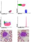
ResultsFlow Cytometry: 93% Double-positive CD4++ CD8+ CD3++ CD2++ CD5++ CD7-/+ CD27(-) CD45RA+ TCR Alpha/Beta+ TCRCBeta1+100% (monoclonal); dim expression of CD56 and CD57; Negative expression: CD25, CD26, CD27, CD28, CD45RO, CD94, CCR7, TCL1, TCR Gamma-Delta (Figure 1).
Morphologyin the analyzed smear, predominance of atypical medium-sized lymphoid cells was observed, with a globose nucleus, generally eccentric, with poorly condensed chromatin with an outline of a nucleolus, and a moderately basophilic, polarized and granular cytoplasm (Figure 1).
References:
- 1.
Shi M, Jevremovic D, Otteson GE, Timm MM, Olteanu H, Horna P. Single antibody detection of T‐cell receptor αβ clonality by flow cytometry rapidly identifies mature T‐cell neoplasms and monotypic small CD8‐positive subsets of uncertain significance. Cytometry B Clin Cytom. 2020;98:99-107.
- 2.
Horna P, Shi M, Jevremovic D, Craig FE, Comfere NI, Olteanu H. Utility of TRBC1 expression in the diagnosis of peripheral blood involvement by cutaneous T-cell lymphoma. J Invest Dermatol. 2021;141:821-829.e2.
- 3.
Alaggio R, Amador C, Anagnostopoulos I, Attygalle AD, Araujo IBO, Berti E, et al. The 5th edition of the World Health Organization classification of haematolymphoid tumours: lymphoid neoplasms. Leukemia. 2022;36:1720-48.
- 4.
Devvit KA, Kern W, Li W, Wang X, Wong AJ, Furtado FM, et al. TRBC1 in flow cytometry: Assay development, validation, and reporting considerations. Cytometry B Clin Cytom. 2024;106:192-202.
- 5.
Horna P, Shi M, Olteanu H, Johansson U. Emerging role of T-cell receptor constant β chain-1 (TRBC1) expression in the flow cytometric diagnosis of T-cell malignancies. Int J Mol Sci. 2021;22:1817.
Figure 1 Flow Cytometry: Double-Positive LGL clone (pink; normal TCD4+ cells in green and normal TCD8+ cells in dark blue). Morphology: atypical medium-sized lymphoid cells was observed, with a globose nucleus, generally eccentric, with poorly condensed chromatin with an outline of a nucleolus, and a moderately basophilic, polarized and granular cytoplasm.


