

Primary myelofibrosis (PMF) is a myeloproliferative neoplasm (MPN) driven by activating mutations in the JAK/STAT pathway. The JAK2 V617F mutation is detected in approximately 60% of patients, CALR indels in 25–30%, and MPL mutations in 5–10%. A subset of patients lacks disease driver mutations and is classified as triple-negative [1]. The mutational landscape of PMF includes other clonal driver mutations affecting genes involved in epigenetic regulation, mRNA splicing, transcriptional regulation, and cell-signaling pathways. Disease-driver and clonal-driver mutations can influence disease presentation and clinical outcomes in patients with PMF, and genomic data are increasingly incorporated into prognostic scores [2–5]. Here, we used a customized target Next Generation Sequencing (NGS) panel to detect mutations in myeloid driver genes in a retrospective real-world series of patients with PMF treated within the public health system in Rio de Janeiro, Brazil. This study aimed to use genomic information to stratify risk and examine its association with disease outcomes.
A total of 32 patients were included, with a median age of 62.5 years at the time of PMF diagnosis (Table S1). Twelve patients (37.5%) had hemoglobin <10 g/dL, 17 (53.1%) had peripheral blood blasts ≥1%, 21 (65.6%) had splenomegaly, and three had a history of thrombosis. At least one constitutional symptom was observed in 12 patients. According to the International Prognostic Scoring System (IPSS), 22% of patients were classified as low risk, 25% as Intermediate-1, 25% as Intermediate-2, and 28% as high risk. Median follow-up time from diagnosis was 5.04 years (95% CI: 2.77–6.73).
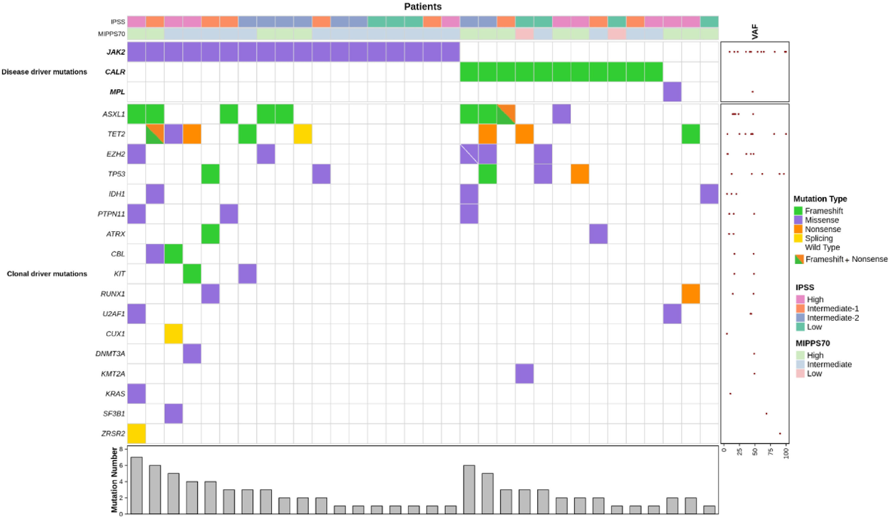
Disease-driver mutations were screened using single-gene approaches, and myeloid clonal driver mutations were detected using NGS (Supplemental material). Among the 32 patients, 14 (43.7%) had DNA samples collected within three months of diagnosis, and the median time from diagnosis to DNA sampling was 7.2 months (Figure S1). A total of 82 mutations were identified across 20 genes (Figure 1 and Table S2). The number of mutations per patient ranged from one to seven, with 22% exhibiting four or more mutations. No significant correlation was found between the number of mutations and the timing of DNA sampling (r = −0.004, p = 0.98). Indeed, clonal evolution in chronic-phase MPN is typically slow, as shown by phylogenetic mutational studies and longitudinal sampling after diagnosis [2,6].

Molecular profile and overall survival in primary myelofibrosis.
A driver mutation was detected in 94% of patients: JAK2 V617F in 54%, CALR indels in 36% (type-1: 24%, type-2: 12%), and MPL W515L in one. Two patients were triple-negative. JAK2 V617F, compared to CALR indels, was associated with higher age, hemoglobin levels, and white blood cell counts (Table S3). Approximately 70% of patients harbored other clonal driver mutations, predominantly frameshift and nonsense mutations affecting ASXL1 and TET2, present in 28% and 25% of patients, respectively. A novel 22 bp deletion in ASXL1 identified by NGS was confirmed as somatic (Figure S2). At least one high-molecular risk (HMR) mutation (ASXL1, EZH2, SRSF2, IDH1/2) [3] was detected in 34% of patients, with five patients presenting two or more HMR mutations. TP53 mutations were found in five patients. Overall, the somatic mutational landscape in our PMF series resembles that reported in larger cohorts [2,4,7].
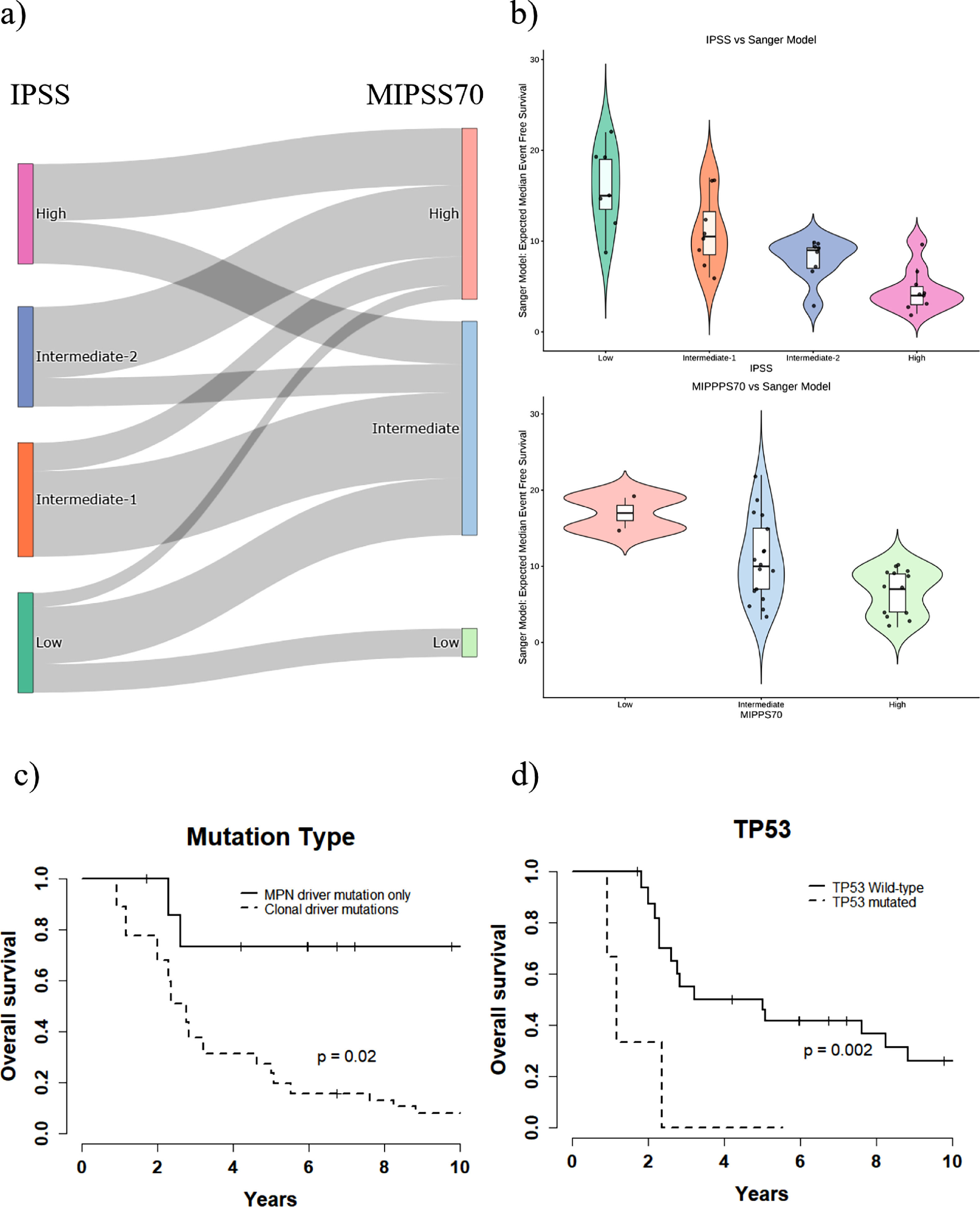
We used mutational and clinical data to assess patient risk according to the Mutation Enhanced International Prognostic Score System (MIPSS70) [5]. MIPSS70 was originally designed for transplant-age patients, and 29 of 32 patients in our series were 70 years old or younger. As MIPSS70 has also demonstrated prognostic utility in all-age PMF cohorts, it was applied to all patients in this study. Compared with IPSS, 37.5% of patients were reclassified into higher-risk categories by MIPSS70. This included 5 of 7 IPPS low-risk patients, and 7 of 16 IPPS intermediate-1/2 patients (Figure 2A). Patients who were upstaged lacked CALR type 1 mutations or harbored HMR mutations or both. Conversely, only 16% of patients were downstaged: 5 of 9 IPSS high-risk patients were reclassified into the MIPSS70 intermediate-risk category. Moreover, we applied the Sanger MPN tool to assess personalized prognosis for each patient [4]. Predicted event free survival ranged from 2–22 years (Figure 2B). These results underscore the importance of integrating NGS molecular analysis to improve risk assessment in a real-world setting.

Risk assessment and overall survival in primary myelofibrosis.
The median overall survival (OS) from diagnosis for all patients was 5.52 years (95% CI: 2.82 to not reached). Because the DNA samples available for NGS were not always from the time of diagnosis, to evaluate the impact of genomic variables and account for potential immortal time bias, all subsequent analyses were calculated from the time of DNA sampling. The median OS from DNA sampling for all patients was 2.82 years (95% CI: 2.29–8.24 years). The number of mutations per patient impacts the OS (Figure S3), and the presence of at least one clonal-driver mutation was associated with shorter OS (Figure 2C). In our series, in contrast to larger studies, no significant difference in survival was observed according to HRM mutation status. This result may be limited by sample size (Figure S3) [3]. While EZH2 mutations were associated with reduced OS, ASXL1 mutations did not significantly affect it (Figure S3). ASXL1 mutations have been associated with worse prognosis in PMF [3]. In contrast to prior reports, the study by Luque Paz et al. found that these mutations alone, in the absence of TP53 or other HRM mutations, did not retain prognostic value [8]. In this report, we found that TP53 mutations were associated with reduced OS (Figure 2D). Indeed, TP53 mutations have been correlated with poorer outcomes and a higher risk of leukemic transformation in PMF [2,4,8–11]. TP53 mutations are often detected at low variant allele frequencies (VAFs) at the time of MPN diagnosis, but clonal evolution can occur as the disease progresses. Leukemic transformation is associated with loss of the second allele and a VAF >50% [2]. Recent data reinforce the prognostic relevance of TP53 multihit mutations in chronic phase myelofibrosis and suggest it should be included in the group of ‘myeloid neoplasms with mutated TP53’ of the International Consensus Classification [10,11]. In our series of PMF patients, 3 of 5 TP53 mutations had VAF >50%. Although this report is based on a retrospective analysis of a small and heterogeneous subset of patients, our data also suggest an adverse impact of TP53 mutations on disease outcome, supporting their inclusion in PMF prognostic scores.
Access to NGS mutational analysis is increasing; however, it is still not widely available in Brazil, particularly within the public health system. Our study demonstrates that molecular profiling offers valuable insights and should be incorporated into the clinical management of patients with PMF.
Ethical approval statementThe study was approved by INCA’s Research Ethics Committee and registered at Plataforma Brasil (CAAE 65,974,117.9. 0000.5274). All patients signed an informed consent form.
Authorship contributionsBMM conceived and designed the study, performed experiments, interpreted data, and wrote the manuscript. IZ conceived and designed the study, interpreted data and contributed to the paper's writing. RRV performed experiments, interpreted the data, prepared figures, and wrote the paper. AHD and CS made the diagnoses, sent patients’ samples, collected clinical and laboratory data, and revised the manuscript. DC, MLR, and MDA performed experiments. MB analyzed genomic data. LJA and JBC analyzed the data, prepared the figures, and contributed to the writing. All authors read and approved the final manuscript.
IZ is a consultant to DASA Genomics. CS has received speaker honorarium and served on the advisory board for Novartis Oncology. All other authors have no conflict of interest to disclose.
This work was supported by grants from PRONON/Ministério da Saúde, FAPERJ, and Coordenação de Aperfeiçoamento de Pessoal de Nível Superior (CAPES)-COFECUB. RRV, MLR, and JBC were supported by grants from the Brazilian National Cancer Institute (Coordenação de Aperfeiçoamento de Pessoal de Nível Superior / Ministério da Saúde).