Journal Information
Vol. 44. Issue 3.
Pages 392-401 (July - September 2022)
Share
Share
Download PDF
More article options
Visits
10083
Vol. 44. Issue 3.
Pages 392-401 (July - September 2022)
Review article
Open Access
Oral manifestations of leukemia as part of early diagnosis
Visits
10083
Reyna Aguilar Quispea,
Corresponding author
reynaaguilarquispe@usp.br

Corresponding author at: Department of Surgery, Stomatology, Pathology and Radiology, Alameda Dr. Otávio Pinheiro Brisola, 9-75, PC: 17012-901. Bauru, São Paulo, Brazil.
, Elizabeth Marques Aguiara, Claudia Teresa de Oliveirab, Ana Cristina Xavier Nevesb, Paulo Sérgio da Silva Santosa
a Faculdade de Odontologia de Bauru da Universidade de São Paulo (FOB USP), Bauru, SP, Brazil
b Hospital Estadual Bauru (HEB) Arnaldo Prado Curvelo, Bauru, SP, Brazil
This item has received

Under a Creative Commons license
Article information
Abstract
Full Text
Bibliography
Download PDF
Statistics
Figures (2)
Abstract
Introduction

The oral cavity can present the first clinical manifestations of leukemia, therefore; it is important to recognize their principal characteristics.

Objective

To identify oral manifestations as the first clinical signs of leukemia.

Methods

This is an integrative review, that gathered data from articles with oral manifestations of leukemia as part of its first clinical features. The were included case reports, case series, clinical research, or reviews with case reports. The variables that were considered relevant: age, sex, sites of the oral lesions, characteristics of the oral lesions, medical history and physical examination, time of evolution, radiographic examination, blood test results, initial diagnosis, differential diagnosis and final diagnosis.

Results

A total of 31 studies were included, with a total of 33 individuals identified. There were 19 (57.57%) males and 14 (42.42%) females. The age range was from 1.6 to 74 years. Acute myeloid leukemia (72.72%) and acute lymphoid leukemia (18.18%) presented more oral manifestations as the first clinical signs of the disease. All individuals with leukemia presented lesions, such as ulcer, erosion, bleeding, ecchymosis, color change of the bluish or pale mucous membranes and areas of tissue necrosis. Hard tissue lesions were less frequent, being 6 (18.18%).

Conclusion

The first clinical manifestations of leukemia can be present in the oral cavity, mainly in acute myeloid leukemia. The principal oral tissues affected were gingival tissue, buccal mucosa and hard and/or soft palate. When hard tissues, such as the maxilla bone or mandible bone were affected, dental mobility was the principal clinical sign.

Keywords:
Leukemia
Diagnosis
Oral
Mouth
Oral manifestations
Full Text
Introduction

Leukemia is a component of oncohematological diseases. Healthy blood cells are replaced with modified immature cells in the bone marrow and circulate through the bloodstream. The classification of lymphoid or myeloid leukemia can be determined by considering the time, whether the leukemia is acute or chronic and the cell type.1,2

The worldwide incidence of leukemia is 437,033 cases, with 30,906 deaths estimated in 2019. The highest prevalence is in males, and acute and chronic leukemia mainly affect the infant/juvenile population and adult population, respectively.3

Given that leukemia is a systemic disease that affects blood components, individuals affected by this disease may present with petechiae, hematomas, ecchymosis and bleeding in different parts of the body, including the mouth, during the physical examination.4

The early identification of this disease allows individuals to start treatment as soon as possible and increases their chances of survival. Therefore, considering that the oral cavity can present the first clinical manifestations of leukemia (MOLs), its recognition from the beginning of the disease is important.5–7

In recent years, few studies have reported oral manifestations as the first clinical signs of leukemia because these manifestations are usually mentioned in clinical case reports that vary from individual to individual and with the type of leukemia.8 This integrative review aimed to identify which tissues of the mouth presented the first MOLs and describes the characteristics that allowed the guiding of the diagnosis of leukemia via clinical signs in the mouth as part of the first clinical manifestations of the disease.

Material and methods

This study is an integrative review that gathered data from articles to describe the clinical MOLs in the oral cavity when they were present as the first clinical signs of the disease and those that contributed to guide the final diagnosis.

Structured research was performed by searching the PubMed and Scopus databases using the following terms: “(first (OR) initial OR early) AND (oral manifestation OR oral sign OR oral lesion) AND (leukemia).”

Articles published in Portuguese, English or Spanish from 2008 to 2020 were included, including case reports, case series, clinical research and reviews that included case reports. The entire publication file had to be available and had to report oral manifestations as the first clinical signs of leukemia. Animal studies, review articles, studies in other languages not mentioned in the inclusion criteria and clinical cases or research articles in which the oral manifestation of leukemia were not part of the first clinical signs of the disease were excluded.

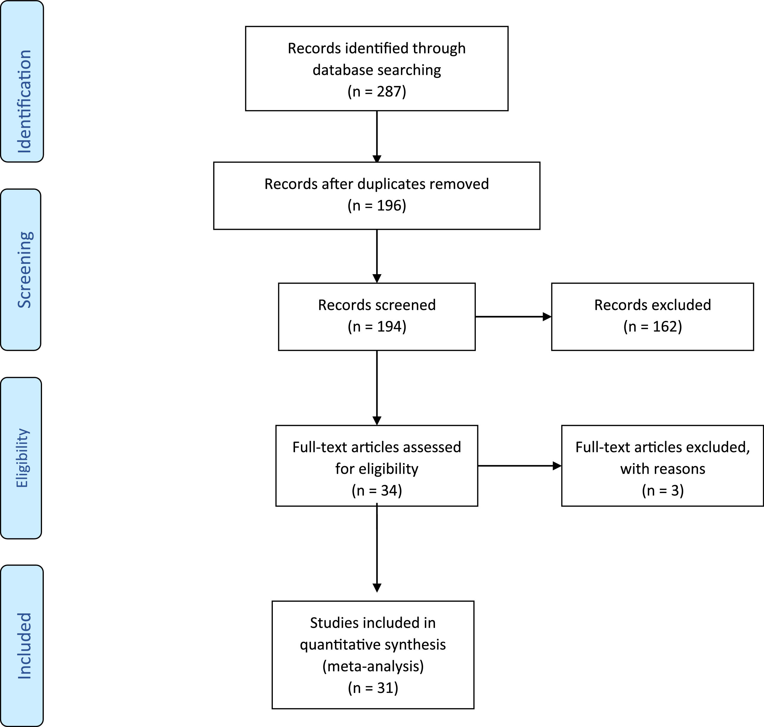
A total of 31 articles fulfilled all the pre-established inclusion criteria. All the information obtained was distributed in a table that contains the variables that were considered relevant: age, sex, sites of the oral lesions, characteristics of the oral lesions, medical history and physical examination, time of evolution, radiographic examination, blood test results, initial diagnosis, differential diagnosis and final diagnosis. (Figure 1)

Figure 1.

Flow diagram of the studies included in the integrative review.

(0.24MB).
Results

A total of 31 studies that mentioned oral manifestations as part of the first clinical signs of leukemia were included. All studies included were clinical case reports or literature reviews that included at least one clinical case report with all the previously described variables.

In the case reports included in this integrative review, the evaluation and initial diagnosis of leukemia were performed principally by dentists who were the first health professionals to come into contact with the patients. Moreover, the dentists also referred the patients to the hematologist and/or oncologist to perform other analyses to establish the final diagnosis. Table 1 describes the characteristics of the 31 case reports.

Table 1.

Principal characteristics of individuals with oral manifestations as first clinical signs of leukemia.

Authors/Year  Gender/Age (years)  Site of oral lesions  Time of evolution  Radiographic exam  Test blood results  Biological reference range  Initial diagnosis  Established differential diagnosis  Final diagnosis (Medical specialist) 
Madhu Singh Ratre, et al., 2018,  M/52  Generalized gingival enlargement  3 months  Panoramic radiography revealed slight to moderate generalized horizontal bone loss (non-signficant for the leukemic diagnosis).  Erythrocyte count 2.68 M (Low). leukocyte count 198,800 (High), neutrophils 0.3%, platelets 30 (Low), blast cells 95%.  Erythrocyte count 3.50 - 5.50 M/μL, leukocyte count 4,000 - 11,000 /μL, neutrophils 40 - 75%, platelets 150 - 450 × 103/μL, blast cells 0%.  Leukemia  None  Acute myeloid leukemia 
Hwa Suk Chae, et.al, 2017  M/12  Bilateral parotid and submandibular glands.  2 days  Panoramic radiograph showed no significant findings.  White blood cells (× 103/μL) 16.24, (× 106 red blood cells (/µL) 3.88, hemoglobin (g/dL) 11.2, hematocrit (%) 31.0, platelets (× 103/μL) 16  White blood cells (× 103/μL) 4.8 - 10.8, red blood cells (/µL) 4.6 - 6.2, hemoglobin (g/dL) 13.0 - 18.0, hematocrit (%) 40.0 - 50.0, platelets (× 103/μL) 130 - 400  Acute sialadenitis  Acute leukemia  Acute lymphoblastic leukemia (B-lymphoid lineage) 
Dalirsani Z, et.al, 2015  M/45  From the right second molar to the left second molar in the mandibular and maxillary bones and hard palate  4 months  The panoramic radiograph: generalized rarefaction of jaw bones, but the inferior alveolar canal was unclear. Thinning of the inferior cortex of the mandible and destruction of the posterior region of the right cortex. Occlusal radiograph: the resorption of the cortical border of the anterior mandible. Periapical radiograph: lamina dura was unclear and indistinct.  White blood cells 53,900/μL, red blood cell 5.3 M/μL, platelet count 319,000/μL; alkaline phosphate 1,530 U/L  Not mentioned  Leukemia or lymphoma  None  Adult T-cell leukemia/ lymphoma 
Guan G, Firth N, 2015  M/49  Palatal gingiva of the maxillary central incisors  2 years  Panoramic and periapical radiographs showed no significant findings.  Hemoglobin (63 g/L), hematocrit (0.19), platelet count (23*109/L) and leukocytes (0.5*109/L)  Hemoglobin 130 – 175 g/L, hematocrit 0.4 – 0.52, platelets 150 – 400 *109 /L, leukocytes 4 – 11 *109 /L  Leukemia  None  Acute myeloid leukemia 
Hasan S, Khan NI, Reddy LB. 2015  F/18  Generalized gingival enlargement  1 month  Panoramic radiography showed no significant findings.  Hemoglobin 7.4 g, red blood cell 1.23 million/cu mm, platelets 60,000/cu mm, white blood cells 812,000/cu mm, neutrophils % 5, lymphocytes % 2  Hemoglobin 11 - 13 g, red blood cells 3.5 - 5.5 million/cu mm, platelets 150,000 - 450,000/cu mm, white blood cells 4,000 - 11,000/cu mm, neutrophils % 40 - 70, lymphocytes % 20 - 40  Leukemia  None  Acute myeloid leukemia 
Babu SP, et.al., 2004  F/43  Generalized gingival enlargment  2 months  Panoramic radiography showed no significant findings.  Platelets: 11,000 150,000 – 400,000, red blood cells: 1.56 × 106, white blood cells: 197,600, neutrophils: 1%, blast cells: 90%  Platelets: 150,000 -400,000, red blood cells: 4.6 - 6.5, white blood cells: 4,000 – 10,000, neutrophils: 45 - 65, lymphocytes: 20 - 40, blast cells: 0  leukemia  Inflammatory enlargement, conditioned enlargement, systemic enlargement and neoplastic enlargement  Acute myeloid leukemia 
Martini V, et.al., 2013  M/52  Mouth floor, labial mucosa and tongue  2 years  None  Normal  Not mentioned  Leukemia  None  Acute lymphoblastic leukemia (mature Natural Killer) 
Gowda TM,et.al., 2013  F/28  Generalized gingival enlargment  2 months  None  White blood cells (cells/cu mm) 48,400, red blood cells (cells/cu mm) 2.8*106, hemoglobin (g/dl) 8.4, hematocrit (%) 25.1, platelets (cells/cu mm) 0.46 lacs, neutrophils (%) 6  White blood cells (cells/cu mm) 4,800 - 10,500, red blood cells (cells/cu mm) 4.6 - 6.2*106, hemoglobin (g/dl)14 - 18, hematocrit (%) 42 - 52, platelets (cells/cu mm) 1.4 - 4.4 lacs, neutrophils (%) 50 - 70  Pregnancy gingival enlargement  None  Acute myeloid leukemia 
Silva BA, et.al., 2012  F/10  The left-nasolabial region  Not mentioned  Occlusal radiograph with no signs of abnormality.  Anemia, leukopenia and thrombocytopenia (there were not reference values),  Not mentioned  Hematopoietic malignancy  None  Chronic lymphocytic leukemia 
Suárez-Cuenca JA, et.al., 2009  M/24  Right lower third molar region  2 days  None  white blood cells 1.5 × 109/L, red blood cells 3.6 × 1012/L, platelets 6.3 × 109/L, hemoglobin 107.0 g/L, hematocrit 0.30,  Not mentioned  Coagulation disorder  Postoperative complication of tooth extraction  Acute myeloid leukemia (subtype M3) 
M Misirlioglu, MZ Adisen, S Yilmaz 2015  M/30  Generalized gingival enlargement  10 days  It was not mentioned what kind of radiograph was made. It revealed no significative changes.  white blood cells 35,000/mm3, platelet 30,000/mm3 and red blood cells 2.7 million/mm3.  Not mentioned  Acute leukemia  Human immunodeficiency virus (HIV)  Acute myeloid leukemia (subtype 4) 
Zacharias Vourexakis, 2015  M/30  Hard palate and the floor of the mouth  3 days  None  5 platelets/nL, 1.1 leukocytes/nL and 22% hematocrit  Not mentioned  Leukemia  None  Acute myeloid leukemia (promyelocityc) 
Mohammed Tag-Adeen, et.al., 2018  F/49  Middle of the soft palate  1 month  The head and neck computed tomography revealed mild bilateral cervical and axillary lymphadenopathy with left maxillary sinus effusion.  Red blood cells: 4.4 × 106, platelets: 274 × 103, leukocytic: 8.3 × 103  Red blood cells: 3.8 – 4.9 × 106, platelets: 158 – 348 × 103, leukocytes: 3.3 – 8.6 × 103 (40% were abnormal)  Candidiasis  Malignant lesion  Adult T-cell Leukemia/Lymphoma 
Chowdhri K, et.al., 2018  M/40  Generalized gingival enlargement involving the buccal, palatal and lingual region  2 months  The panoramic radiograph revealed generalized horizontal bone loss.  White blood cell (× 103/μL) 44.78, red blood cells (× 103/μL) 1.47 4.00 - 6.00, hemoglobin (g/dL) 4.9 13.5 - 17.5, hematocrit (%) 14.5, platelet (× 103/μL) 65  White blood cell (× 103/μL) 4.00 - 11.00, red blood cells (× 103/μL) 4.00 - 6.00, hemoglobin (g/dL) 5 - 17.5, hematocrit (%) 42 - 52, platelet (× 103/μL) 150 - 450  Acute leukemia  Acute manifestaion of systemic disease  Acute monocytic leukemia (subtype M5b) 
M.Alessandriniaet,et al., 2012  F/74  The upper left vestibule next to the left upper central incisor and upper left secondary premolar  Not mentioned  Computed tomography confirmed a tissue-like mass, 3.2 × 0.7 × 0.3 cm located in the left vestibule of the maxillary bone.  17,300 platelets/mL, 12,300, white blood cells/mL with 51% of lymphocytes and 4.1 million, red blood cells/mL.  Not mentioned  Squamous cell carcinoma, deep fungal infections  Leukemia  Chronic lymphocytic leukemia 
A. Chatzipantelis, P. A. Atkin, 2018  M/50  Gingiva adjacent to the upper right first molar and buccal right mandibular buccal sulcus  1 week  The panoramic radiograph had no significant findings.  White blood cell count 0.5, hemoglobin 103, platelet count 21, red blood cell count 3.31, hematocrit 0.3, neutrophil count 0.1  White blood cell count 74.4 1.3 0.5 4 – 11 × 10(9)/L, hemoglobin 130 – 180 g/L, platelet count 150 – 400 × 10(9)/L, red blood cell count 4.50 – 6.00 × 10(12)/L, hematocrit 0.40 – 0.52 L/L, neutrophil count 1.7 – 7.5 10(9)/L  Acute leukemia  None  Acute myeloid leukemia 
Shimizu R, et.al., 2017  M/12  The right maxillary molar and the second primary molar  1 month  The panorama: diffuse opacification in the right maxillary sinus cavity, non-contrast computed tomography showing osteoblastic lesions of the anterior and posterior walls of the maxillary sinus and thickening of the surrounding bone and soft tissue of the right maxilla were observed.  Mild leukopenia and high serum alkaline phosphatase  Not mentioned  Osteosarcoma of the right maxilla  Osteogenic sarcoma, Ewing's sarcoma, and hematologic malignancy or metastatic malignancies  Acute myeloid leukemia (subtype M5a) 
Zhang Y, et.al., 2010  F/9  Right facial asymmetry,the maxillofacial region clearly protruding outward.  2 weeks  A contrast computed tomography scan of the maxillae demonstrated a homogeneous mass located in the right maxillary fossa extending backward into the infratemporal fossa without bone destruction.  White blood cell count of 31.6 × 109/L, a platelet count of 272 × 109/L, and hemoglobin and red blood cell count of 107 g/L and 3.87 × 1012/L,  Not metioned  Leukemia infiltration  None  Acute lymphoid leukemia (High-risk B-cells) 
Mattheos K. et.al., 2010  F/70  The alveolar socket of the extracted left mandibular first premolar  20 days  The radiographic examination of the mandible did not show any bone damage.  Not mentioned  Not mentioned  Myeloid sarcoma (biopsia)  None  Acute myeloid leukemia 
Chung SW, et.al., 2011  M/35  The lower anterior and left posterior teeth  4 months  A panoramic radiograph revealed an osteolytic lesion on the left mandibular body and ascending ramus area with severe vertical bone loss on the lower left posterior mandible. The computed tomography showed an osteolytic lesion on the entire left half of the mandible.  Red blood cells 3.93 × 106/ml), but normal white blood cell count (10.39 × 103/ml), platelet count (261 × 103/ml)  Not menioned  Hematologic malignancy  Acute osteomyelitis of the mandible, osteosarcoma  Acute lymphoid leukemia (Burkitt type) 
Sharon Aronovich, Thomas W. Connolly, 2008  M18  Mandibular left third molar  1 week  A panoramic radiograph showed a 30° mesial tilt of the mandibular left third molar with minor ovate radiolucencies mesial to roots of the mandibular left third molar and the mandibular right third molar. These radiographic findings were deemed inconclusive.  Blood count showed 44% lymphoblasts with profound neutropenia and severe thrombocytopenia (17,000 platelets/mm3),  Not mentioned  Pericoronitis with occlusal trauma  None  Acute lymphoblastic leukemia 
Sepúlveda E, et.al., 2012  M/6  Gingiva and hard palate  2 weeks  None  hemoglobin 7.2 (g/dl), hematocrit 20 (%), white blood cells 26,600 (cells/tnl^, neutrophil absolute count 530 (cells/ml'), lymphocyte absolute count 14,900 (cells/ml^, platelets 20,000 (cells/mP)  Hemoglobin (g/dl) hematocrit (%) white blood cells (cells/tnl^ neutrophil absolute count (cells/ml') Lymphocyte absolute count (cells/ml^ platelets (cells/mP) 0 CONFIRM DIAGNOSIS  Leukemia  Complication of hemorrhage after tooth extraction  Acute myeloid leukemia (subtype M3) 
Preeti Chawla Arora, et.al., 2020  F/18  Several ulcers on the tip of the tongue, buccal mucosa and gingiva; mild enlargement of interdental gingiva in maxillary and mandibular anterior teeth  One week  None  Hemoglobin 7.8 g%, high leukocyte count 34,900 mm3 and low platelet count 25,000 mm3  Not mentioned  Aphthous stomatitis and HIV-associated oral ulcers  None  Acute myeloid leukemia 
  M/25  Generalized gingival enlargement  One month  None  hemoglobin level (5 g %), leukocyte count 65,000 mm3, platelet count 25,000 mm3 and red blood count 1.69 million mm3  Not mentioned  Gingival enlargement induced by drugs  Gingival enlargement due to leukemia  Acute myeloid leukemia 
Mahnaz Fatahzadeh and A. Michael Krakow, 2008  M/26  Anterior gingival enlargement  3 days  It was not mentioned what kind of radiograph was made. It revealed no significative changes.  White blood count: 112,000 cells/mL3; platelets 15,000 cells/mL3; hemoglobin: 6.6 g/dL  White blood count: normal/nl 5,000 –10,000; platelets: 150,000 – 400,000; hemoglobin:13.8 – 17.2 g/dl  Generalized gingivitis, acute herpetic gingivostomatitis  Systemic disorders of hematological, immunosuppressive and infectious etiology  Acute myeloid leukemia 
Y-W Fu, H-Z Xu, 2017  F/27  Generalized gingival enlargement  3 months  None  Blood cell count of 9.68 9 109/L, with a hemoglobin count of 64.0 g/L and a platelet count of 17 9 109/L,  Not mentioned  Gingival enlargement in pregnancy  none  Acute myeloid leukemia (monocytic lineage) 
Hyun-Chang Lim, Chang-Sung Kim, 2014  F/56  Generalized gingival enlargement  20 days  Panoramic radiograph revealed generalized horizontal bone loss that was most prominent in the anterior maxillary region.  white blood count (71.52 × 103/μL), decreased red blood cell (2.14 × 106/μL) and platelet (83 × 103/μL) counts,  Not mentioned  Manifestation of underlying systemic disease  None  Acute myeloid leukemia (subtype M4) 
  F/49  Generalized gingival enlargment  3 weeks  Panoramic radiograph and computed tomography revealed severe periodontal destruction.  white blood count of 38.01 × 103/μL and decreased red blood cell (1.46 × 106/μL) and platelet (20 × 103/μL) counts.  Not mentioned  Complications and abscess after tooth extraction  None  Acute myeloid leukemia (subtype M4) 
Srinivas Rao Ponnam, et.al., 2014  F/45  Gingival enlargement in the anterior region of the upper jaw and  1 month  Panoramic radiograph opacification of the left maxillary sinus and presence of root stumps in relation to 21st, 22nd and 25th teeth  Not mentioned  Not mentioned  Metastatic tumor of unknown origin  Small round cell tumor (after biopsy)  Acute myeloid leukemia 
Mechery Reenesh, Singh Munishwar, Saroj Kumar Rath, 2012  M/32  generalized gingival enlargement. Ecchymosis in the floor of the mouth  5 months  None  Hemoglobin 5.6 gm%; red blood count 1.41 million/cu mm, platelets 25,000/cu mm; white blood cell count 112,000/cu mm  Hemoglobin 11.5 - 16.5 gm%; 3.5 - 6.0 million/cu mm; 1,50,000 - 4,50,000/cu mm; 4,000 - 11,000/cu mm  Oral manifestation of systemic disease  None  Acute myeloid leukemia (subtype M4) 
Bianca Piscinato Piedade Rosa, et.al., 2018  M/47  Ecchymoses in the left ventral surface of the tongue and a hematoma in the gingiva around the mandibular left canine and first premolar  1 month  None  Red blood count 3.35 million/mm³, platelets 22,000/mm³, leukocytes 67,200/mm³  Red blood count 4.3 to 6.1 million/mm³; 150,000 to 450,000/Mm³; leukocytes 3,500 to 11,000/Mm³  Acute leukemia  None  Acute myeloid leukemia (hypogranular variant) 
Paulo Sérgio da Silva Santos, et.al., 2010  M/43  Generalized gingival enlargement  15 days  Panoramic radiograph had no significant findings  Hemoglobin, 10.5 g/dL; hematocrit, 30.3 percent; leukocytes, 153,000/L with 13 percent of blasts; platelets, 73,000/L;  Not mentioned  Necrotizing acute gingivitis  Opportunistic infection caused by AIDS; extramedullary leukemic infiltrate  Acute myeloid leukemia (subtype M5) 
Cristina Vázquez-Martínez, et.al., 2018  F/1.6  Inflammatory lesion in the right mandibular body  4 days  Ultrasound of soft tissue revealed hypoechogenic mass of 13 mm in the right mandibular body, probably related to a dental germ  white blood cell count 165,000/mm³; platelets 95,000/mm³; hemoglobin 11.7 g/dl  Not mentioned  Cyst of dental eruption  Myeloid sarcoma (biopsy)  Acute myeloid leukemia (subtype Mab) 

A total of 33 individuals were identified in the selected studies: 19 males (57.57%)9,10,11–18,19–22 and 14 females (42.42%).23–30 The age ranged from 6 to 74 years and only 6 individuals (18.18%) were between 1.6 and 12 years old.10,19,22,26,31,32

Among the types of leukemia, oral manifestations were present principally in acute myeloid leukemia (24 individuals [72.72%]),9,12,14–19,22,23,24,25,30,32 followed by acute lymphoid leukemia (6 individuals [18.18%]),10,13,20,21,26,31 adult T-cell leukemia/lymphoma (2 individuals [6.06%]) and chronic lymphoid leukemia (1 individual [3.03%]).
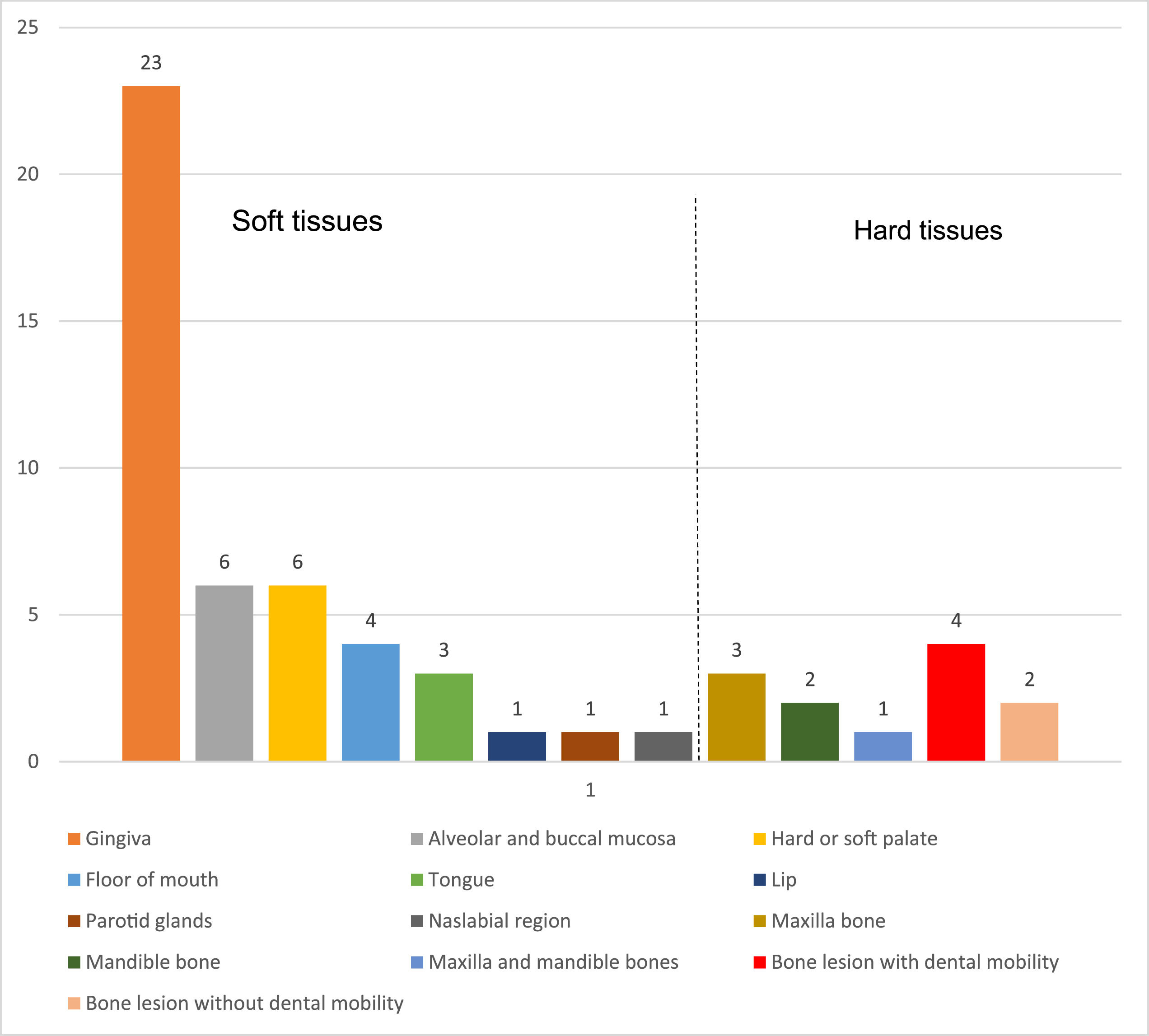
First clinical MOL in oral tissues

Oral MOLs as the first clinical signs were more common in soft tissues (27 individuals [81.81%]) than those in hard tissues (6 individuals [18.18%]).11,19,20,28,31,32Figure 2 shows the different sites.

Soft tissues

The oral MOLs were in gingival tissue (63.63%),9,12,17–19,21,23–25,26,27,30,33–39,15,40 alveolar and jugal mucosa (18.18%)14,18,21,22,26,28 and hard or soft palate (18.18%).9,11,15,16,27,30 Other regions, such as the floor of the mouth (12.12%),13,16,23,38 tongue (9.09%),13,33,39 lip (3.03%),13 parotid glands (3.03%),10 and nasolabial region (3.03%),26 were less frequent.

The main lesions found in the soft tissues of the mouth were ulcer, erosion, bleeding, ecchymosis, color change of the bluish or pale mucous membranes, and areas of tissue necrosis. Among the case reports, 13 individuals (39.99%) reported pain as the principal symptom,9,13,28,30,31,14,15,17–21,25 which was also principally spontaneous.

Figure 2.

Site of first clinical signs of leukemia in oral cavity.

(0.2MB).
Hard tissues

Among the oral MOLs in hard tissues, two cases corresponded to ALL,20,31 two cases corresponded to AML,19,32 one case corresponded to chronic lymphocytic leukemia28 and one case corresponded to adult T-cell leukemia/lymphoma.11 Dental mobility was the most common clinical condition when hard tissues were affected. Panoramic radiography and computed tomography were used to evaluate the bone structures. The main characteristics of the radiographic images were cortical expansion of the alveolar bone, osteolytic areas, thickening of the bone surrounding the teeth with mobility and severe vertical bone loss.

Differential diagnosis of the oral MOLs

In soft tissues, the differential diagnoses were gingival enlargement during pregnancy,25,35 gingival enlargement induced by drugs,41 squamous cell carcinoma and deep fungal infection,28 pericoronarite,21 oral manifestation of HIV,15,33 postoperative complications of extraction,14,36 candidiasis,27 necrotizing acute gingivitis,40 dental cyst eruption32 and herpetic gingivostomatitis.34 In addition, when the oral manifestation was in the maxillary bones, the presumptive diagnoses were acute osteomyelitis,20 osteosarcoma19,20 and Ewing sarcoma.19

General clinical signs and symptoms of leukemia

The general signs and symptoms, in addition to the oral manifestations, were mainly the following: weakness, fatigue, lethargy or tiredness in 11 individuals (45.45%);9,12,15,17,18,20,23,26,30,36 lymphadenopathies in 8 individuals (36.36%);9,14,15,17,22,25–27 mucosal pallor in 6 individuals (27.27%);12,14,15,22,26 weight loss in 4 individuals (18.18%)15,17,23,38 and loss of appetite in 5 individuals (18.18%).9,16,23,26,32 Adjacent areas, such as the head and neck regions, presented submandibular lymphadenopathy,9,10,15,26,34 cervical lymphadenopathy,17,27,34 dysphagia,9,16,17 facial asymmetry10,26,31,37 and trismus.10,14

Other clinical signs and symptoms that were present, but were less frequent, included genital ulcers,13 vomiting,11 nausea,11,17 diarrhea,11 dizziness,17 abdominal swelling,12 petechiae in the leg and forearm,12,21 musculoskeletal pain,26 increased volume in the pre-sternal region26 and chills and joint pain.21

Results of laboratory tests that led to the presumptive diagnosis of oral MOLs

The blood count was the main blood test requested to help support the presumptive diagnosis of an oral MOL. Thrombocytopenia was present in 22 cases (59.09%),9,10,24,25,31–38,12,39,40,14,15,17,18,21–23 anemia was present in 23 cases (63.63%),9,10,25,26,31–38,12,39,40,14,15,17,20,22–24 leukocytosis was present in 19 cases (45.45%)9,11,31–40,13,15,17,18,22–25 and leukopenia was present in 5 cases (22.72%).10,12,14,19,26

Discussion

The diagnosis of leukemia is based initially on clinical signs and symptoms that allow for the initial diagnosis of the disease. However, the definitive diagnosis is made by different complementary tests, such as karyotyping, flow cytometry and bone marrow biopsy.6,42 Early diagnosis allows the individual to start antineoplastic treatment as soon as possible, thus increasing the chance of survival and improving the prognosis.43

Before the diagnosis of leukemia, individuals can present with fatigue, fever, adenomegaly, hepatosplenomegaly, persistent or recurrent infections, hematomas, pallor, petechiae and unexpected bleeding from the skin and mucous membranes, including the oral mucosa.6,43 These symptoms and clinical signs may be similar and appear in other systemic diseases, thus hindering an early diagnosis.2 In the current review, the oral MOLs were the main complaint during examination, however, fatigue, lethargy or tiredness, lymphadenopathies, pallor, weight loss and loss of appetite were also reported, as well as the presence of dysphagia, facial asymmetry and trismus.

In this review, among the different types of leukemia with oral manifestations as the first clinical signs, acute leukemia was the most common, particularly acute myeloid leukemia; this finding is similar to that of previous studies.5,44,45 The first oral MOLs include gingival bleeding, gingival hyperplasia, ulcers and petechiae.4,5,44–46 When oral MOLs are present, it is easier for patients to identify these clinical signs because of the visibility of the mouth, compared to other body structures. All cases that were included in this review reported oral lesions as the main complaint, which was the principal reason for visiting dentists before being referred to specialists in the field of oncohematology for the final diagnosis of leukemia.

Oral MOLs can be found mainly in the gingiva, lips, hard and/or soft palate and tongue.5 In all case reports in this review, the clinical signs of leukemia in the mouth affected mainly the soft tissues. The gingival tissue had the highest manifestation9,12,26,27,30,15,17–19,21,23–25 among the types of leukemia and the mouth floor, parotid, tongue and nasolabial region10,13,16,23,26 had the lowest manifestations. Some cases reported spontaneous pain at the time of the complaint and this was one of the main reasons for seeking dental care.

In this review, it was identified that bone structures, such as the maxilla and/or mandible, had the principal features of areas with increased volume, rapid growth and dental mobility without apparent cause as part of the first clinical signs of leukemia. These characteristics led to the suspicion of malignant bone lesions, thus necessitating evaluation by imaging exams.11,19,20,28,31

Complementary imaging examinations, such as panoramic radiography and computed tomography, are essential to evaluate bone structures in greater detail. For this reason, when radiography revealed the characteristics of malignant bone lesions, the patients were referred immediately to specialists in hematology/oncology.19,20

Other dentists used the radiographs to guide the incisional biopsy, which was also useful for early referral to a specialist.28,31 Dentists can perform a biopsy if the patient agrees with the procedure. Among all the case reports of this review, one patient refused biopsy. Nevertheless, because of the malignant characteristics, the patient was immediately referred to a specialist.11 Therefore, malignant characteristics in radiography, with clinical signs and symptoms that are suspected of being oral MOLs, need to be immediately referred to a specialist in hematology/oncology, regardless of whether an oral biopsy needs to be performed.

Bone alterations as oral MOLs are rare, however, when present, they can provoke facial asymmetry, which can be the main reason for seeking dental care.31 Some bone alterations were also present in children.19,31 Therefore, bone alterations can also appear in patients who are still undergoing bone development.

The literature mentioned that children and adolescents with leukemia, who are undergoing bone development and bone maturation, respectively, had bone areas with osteotropic characteristics that can promote the local invasion of malignant cells into bone structures, thus provoking their destruction.47,48

Moreover, hypercalcemia was found in adult patients with bone MOLs.11,36 One of the clinical reports in this review mentioned that hypercalcemia was associated with the ectopic production of the parathyroid hormone protein, in addition to a severe increase in alkaline phosphatase levels.11 Oral manifestations in bone structures as the first clinical signs of leukemia are considered extremely rare, however, after chemotherapy, the bone lesions can regress and the follow-up can be performed via radiography examination.11,19,20,28,31

The diagnosis of leukemia can be challenging and complex, particularly when the oral MOLs have characteristics similar to those of other systemic diseases and specific oral diseases. In this review, 11 cases of oral MOLs were initially confused with other lesions that presented clinical signs and symptoms that are identical to those of some oral diseases.14,15,35,36,19–21,25,27,28,32,33

For example, a case report included in this review mentioned pericoronitis as an initial diagnosis, which is characterized by inflammation, pain, trismus and sometimes, infection associated with the periodontitis eruption of the third molar tooth.21 The oral MOL in this case presented characteristics that initially mimicked the oral MOL. Therefore, the referral to the hematologist was delayed. Another case report in which the final diagnosis was leukemia had pregnancy gingivitis or hyperplasia as the first diagnosis.25 Some pregnant patients with relative frequency may present with changes in the gingival tissue, such as gingivitis. Therefore, considering that the patient was pregnant, the initial diagnosis coincided with the clinical condition of the patient.

Complementary tests, such as the complete blood count, were essential in establishing the initial diagnosis based on the first oral MOL. In this review, a blood count was requested for all patients and this approach allowed for the immediate referral to specialists in hematology/oncology. The main alterations were leukocytosis, thrombocytopenia, anemia and the presence of blasts in the blood count, which was essential and useful, not only in diagnosing leukemia, but also in evaluating the prognosis and guiding the treatment of the patient. An early diagnosis of leukemia corresponds to a high chance of survival.

This integrative review included data from patients of different ages and therefore, we did not analyze the data by age group. This was a limitation because there is a possibility of finding specific first clinical signs of leukemia in the oral cavity of children, young people and the elderly.

Conclusions

Leukemia may present with oral manifestations as part of its first clinical signs. Acute leukemia was the type with the most oral manifestations, particularly acute myeloid leukemia. The anatomical structures most affected in the oral cavity were the gingival tissue, jugal mucosa and hard or soft palate. When oral MOLs were present in hard tissues, such as the maxilla or mandible, dental mobility was the main clinical characteristic. The main complementary test to help formulate the initial diagnosis of leukemia and referral to specialists, such as a hematologist and/or oncologist, was the complete blood count, which can identify leukocytosis, anemia, and thrombocytopenia.

Funding

This study was financed in part by the Coordenação de Aperfeiçoamento de Pessoal de Nível Superior - Brasil (CAPES) - Finance Code 001.

References
[1]
J Ferlay, M Colombet, I Soerjomataram, C Mathers, DM Parkin, M Piñeros, A Znaor, F. Bray.
Estimating the global cancer incidence and mortality in 2018: GLOBOCAN sources and methods.
Int J Cancer, 144 (2019), pp. 1941-1953
[2]
A Redaelli, JM Stephens, BL Laskin, CL Pashos, MF. Botteman.
The burden and outcomes associated with four leukemias: AML, ALL, CLL and CML.
Expert Rev Anticancer Ther, 3 (2003), pp. 311-329
[3]
F Bray, J Ferlay, I. Soerjomataram.
Global Cancer Statistics 2018 : GLOBOCAN estimates of incidence and mortality worldwide for 36 cancers in 185 countries.
CA Cancer J Clin, 68 (2018), pp. 394-424
[4]
E Watson, RE Wood, WG Maxymiw, AD Schimmer.
Prevalence of oral lesions in and dental needs of patients with newly diagnosed acute leukemia.
J Am Dent Assoc, 149 (2018), pp. 470-480
[5]
GL Hou, JS Huang, CC. Tsai.
Analysis of oral manifestations of leukemia: a retrospective study.
[6]
VP Burke, JM Startzell.
The Lleukemias. Oral Maxillofac Surg Clin North Am, 20 (2008), pp. 597-608
[7]
F Cammarata-Scalisi, K Girardi, L Strocchio, P Merli, A Garret-Bernardin, A Galeotti, et al.
Oral manifestations and complications in childhood acute myeloid leukemia.
Cancers (Basel), 12 (2020), pp. 1634
[8]
CF Francisconi, RJ Caldas, LJ Martins, CM Rubira.
Santos PS da S. Leukemic oral manifestations and their management.
Asian Pacific J Cancer Prev, 17 (2016), pp. 911-915
[9]
M Ratre, R Gulati, S Khetarpal, A. Parihar.
Regular oral screening and vigilance: can it be a potential lifesaver?.
J Indian Soc Periodontol, 22 (2018), pp. 171
[10]
HS Chae, J-S Byun, J-K Jung, J-K Choi.
Trismus as an orofacial manifestation of acute lymphoblastic leukemia.
J Oral Med Pain, 42 (2017), pp. 49-52
[11]
R Shimizu, N Ohga, M Miyakoshi, T Asaka, J Sato, Y. Kitagawa.
Unusual maxillary osteoblastic and osteolytic lesions presenting as an initial manifestation of childhood acute myeloid leukemia: a case report.
Quintessence Int (Berl), 48 (2017), pp. 149-153
[12]
SW Chung, S Kim, JR Choi, TH Yoo, IH. Cha.
Osteolytic mandible presenting as an initial manifestation of an adult acute lymphoblastic leukaemia.
Int J Oral Maxillofac Surg, 40 (2011), pp. 1438-1440
[13]
S Aronovich, TW. Connolly.
Pericoronitis as an initial manifestation of acute lymphoblastic leukemia: a case report.
J Oral Maxillofac Surg, 66 (2008), pp. 804-808
[14]
E Sepúlveda, U Brethauer, E Fernández, G Cortés, C. Mardones.
Oral manifestations as first clinical sign of acute myeloid leukemia: report of a case.
Pediatr Dent, 34 (2021), pp. 418-421
[15]
Z Dalirsani, A Javadzade Bolouri, Z Delavarian, S Bidad, M Sanatkhani, M Amirchaghmaghi.
Human T-lymphotropic virus-1 associated with adult T-cell lymphoma/leukemia and generalized expansion of palatal and jaw bones: a rare case report.
J Dent (Shiraz Univ Med Sci, 16 (2015), pp. 214-218
[16]
Guan G, Firth N. Oral manifestations as an early clinical sign of acute myeloid leukaemia : a case report. 2015:123-7. doi:10.1111/adj.12272.
[17]
V Martini, P Schiavone, R Bonacina, U Mariani, A. Rambaldi.
NK leukemia: a rare case of oral manifestations representing the initial sign.
Ann Stomatol (Roma), 4 (2013),
[18]
JA Suárez-Cuenca, JL Arellano-Snchez, AA Sácherling-Ocampo, G Sánchez-Hernández, D Pérez-Guevara, JR. Chalapud-Revelo.
Rapidly progressing, fatal and acute promyelocytic leukaemia that initially manifested as a painful third molar: a case report.
J Med Case Rep, 3 (2009), pp. 1-6
[19]
M Misirlioglu, MZ Adisen, S. Yilmaz.
Diagnosis of acute myeloid leukemia in a dental hospital; Report of a case with severe gingival hypertrophy.
Niger J Clin Pract, 18 (2015), pp. 573-576
[20]
Z. Vourexakis.
Oral lesions presenting as an early sign of acute leukaemia.
BMJ Case Rep, 2015 (2015), pp. 2014-2015
[21]
K Chowdhri, S Tandon, A Lamba, F. Faraz.
Leukemic gingival enlargement: a case report and review of literature.
J Oral Maxillofac Pathol, 22 (2018), pp. 77
[22]
A Chatzipantelis, PA. Atkin.
Intra-oral noma-like lesion associated with acute myeloid leukaemia: a case report.
Oral Surg, 11 (2018), pp. 306-310
[23]
S Hasan, N Khan, Reddy Lb.
Leukemic gingival enlargement: report of a rare case with review of literature.
Int J Appl Basic Med Res, 5 (2015), pp. 65
[24]
SPKK Babu, V Kashyap, P Sivaranjani, S. Agila.
An undiagnosed case of acute myeloid leukemia.
J Indian Soc Periodontol, 18 (2014), pp. 95-97
[25]
TM Gowda, R Thomas, SM Shanmukhappa, G Agarwal, DS. Mehta.
Gingival enlargement as an early diagnostic indicator in therapy-related acute myeloid leukemia: a rare case report and review of literature.
J Indian Soc Periodontol, 17 (2013), pp. 248-252
[26]
BA Silva, CR Siqueira, PH Castro, SS Araújo, LE. Volpato.
Oral manifestations leading to the diagnosis of acute lymphoblastic leukemia in a young girl.
J Indian Soc Pedod Prev Dent, 30 (2012), pp. 166-168
[27]
M Tag-Adeen, K Hashiguchi, Y Akazawa, K Ohnita, S Yasushi, N Daisuke, et al.
An unusual presentation of adult T-cell leukemia/lymphoma.
Ecancermedicalscience, 12 (2018), pp. 1-7
[28]
M Alessandrini, A Micarelli, F Mugnaini, A De Padova, I Pavone, E Bruno.
Unusual case of oral chronic lymphocytic leukemia presenting as recurrent epistaxis and asymptomatic intraoral swelling.
Rev Stomatol Chir Maxillofac, 113 (2012), pp. 455-457
[29]
R Zhang, Y Teng, L Zhu, J Lin, X Yang, G Yang, et al.
Odontoblast β-catenin signaling regulates fenestration of mouse Hertwig's epithelial root sheath.
Sci China Life Sci, 58 (2015), pp. 876-881
[30]
MK Papamanthos, AE Kolokotronis, HE Skulakis, AM Fericean, MT Zorba, AT Matiakis.
Acute myeloid leukaemia diagnosed by intra-oral myeloid sarcoma. A case report.
Head Neck Pathol, 4 (2010), pp. 132-135
[31]
Y Zhang, L Li, L Ma, F Zhou, L Liu.
Maxillofacial mass as the first presentation of acute lymphoblastic leukemia in a nine-year-old girl.
Auris Nasus Larynx, 37 (2010), pp. 377-380
[32]
C Vázquez-Martínez, M Redondo-Alamillos, AI Romance-García, G. Sánchez-Aniceto.
Sarcoma mieloide intraoral: Forma poco común de debut de la leucemia aguda mieloide.
Rev Esp Cir Oral y Maxilofac, 41 (2019), pp. 41-43
[33]
PC Arora, A Arora, S. Arora.
Oral manifestations as an early clinical sign of acute myeloid leukemia: a report of two cases.
Indian J Dermatol, 65 (2020), pp. 241-243
[34]
M Fatahzadeh, AM. Krakow.
Manifestation of acute monocytic leukemia in the oral cavity: a case report.
Spec Care Dent, 28 (2008), pp. 190-194
[35]
YW Fu, HZ. Xu.
Gingival enlargement in a pregnant woman with acute monocytic leukaemia: a case report.
Aust Dent J, 62 (2017), pp. 386-389
[36]
HC Lim, CS. Kim.
Oral signs of acute leukemia for early detection.
J Periodontal Implant Sci, 44 (2014), pp. 293-299
[37]
SR Ponnam, G Srivastava, N Jampani, V. Kamath.
A fatal case of rapid gingival enlargement: case report with brief review.
J Oral Maxillofac Pathol, 18 (2014), pp. 121-126
[38]
M Reenesh, S Munishwar, SK. Rath.
Generalised leukaemic gingival enlargement: a case report.
J Oral Maxillofac Res, 3 (2012), pp. 1-6
[39]
BP Piedade Rosa, FA Ito, FC Trigo, LT Mizuno, AT Junior.
Oral manifestation as the main sign of an advanced stage acute promyelocytic leukemia.
Acta Stomatol Croat, 52 (2018), pp. 358-362
[40]
Da Silva Santos PS, A Fontes, A Flávio De, SCOMH De Sousa.
Gingival leukemic infiltration as the first manifestation of acute myeloid leukemia.
Otolaryngol - Head Neck Surg, 143 (2010), pp. 465-466
[41]
SG Hsieh, S Hibbert, P Shaw, V Ahern, M. Arora.
Association of cyclophosphamide use with dental developmental defects and salivary gland dysfunction in recipients of childhood antineoplastic therapy.
Cancer, 117 (2011), pp. 2219-2227
[42]
J Zawitkowska, M Lejman, A Zaucha-Prażmo, K Drabko, M Płonowski, J Bulsa, et al.
Clinical characteristics and analysis of treatment result in children with Ph-positive acute lymphoblastic leukaemia in Poland between 2005 and 2017.
Eur J Haematol, 101 (2018), pp. 542-548
[43]
DA Arber, A Orazi, R Hasserjian, J Thiele, MJ Borowitz, MM Le Beau, et al.
The 2016 revision to the World Health Organization classification of myeloid neoplasms and acute leukemia.
Blood, 127 (2016), pp. 2391-2405
[44]
N López-Valverde, A López-Valverde, RG Diego, JM de, Ramírez, J Flores-Fraile, J. Muriel-Fernández.
Gingival hyperplasia as an early manifestation of acute myeloid leukemia. A retrospective review.
J Clin Exp Dent, 11 (2019), pp. 1139-1142
[45]
R Busjan, J Hasenkamp, G Schmalz, R Haak, L Trümper, D. Ziebolz.
Oral health status in adult patients with newly diagnosed acute leukemia.
Clin Oral Investig, 22 (2018), pp. 411-418
[46]
AO Gomes, A Silva Junior, CW Noce, M Ferreira, A Maiolino, SR Torres.
The frequency of oral conditions detected in hematology inpatients.
Hematol Transfus Cell Ther, 40 (2018), pp. 240-244
[47]
A Schajnovitz, DT. Scadden.
Bone ’ s dark side : mutated osteoblasts implicated in leukemia.
Nat Publ Gr, (2014), pp. 383-384
[48]
Recine M, Castellano-sanchez AA, Sheldon J. Precursor B-cell lymphoblastic lymphoma /leukemia presenting as osteoblastic bone lesions. 2002;6(4):236-243. doi:10.1053/adpa.2002.34733.
Idiomas
Hematology, Transfusion and Cell Therapy
Article options
Tools